The prevention, assessment and management of skin tears
Jackie Stephen-Haynes
Jackie Stephen-Haynes
RGN, DN, DipH, BSc (Hons), ANP. Professor in Tissue Viability and Consultant Nurse
Professional Development Unit, Birmingham City University and Worcestershire Health and Care NHS Trust
Michelle Deeth
RGN, MSc
Honorary Tissue Viability Nurse
Worcestershire Health and Care NHS Trust
Skin tears are under-reported and often poorly managed, but cause pain and decreased quality of life for patients but increasing incidence among our ageing patient population means they are likely to become one of the largest problem areas in wound care
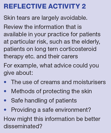
Skin tears are increasingly recognised as a mostly avoidable, yet common, acute wound in patients with fragile skin, including both the elderly and the newborn. It has been suggested that skin tears will become one of the largest problem areas in wound care. It is therefore important to focus on prevention: creating a safe environment, identifying and, where possible, removing those things that can cause skin tears, particularly in the elderly population.
Lower leg skin tears, if not managed appropriately, have the potential to ulcerate. Skin tears may, indeed, increase both patient morbidity and mortality.1 While skin tears are under-reported and may be poorly assessed and managed, patients with them describe increased pain and decreased quality of life.2
This article covers:
- Key aspects of the physiology of the skin and the development of skin tears
- The importance of appropriate management options, with an analysis of the current literature regarding wound dressings and skin tears
- The need for prompt and appropriate categorisation of a skin tear, to enable the clinician to prevent further pain and reduce the likelihood of progression to ulceration and/or cellulitis.
ANATOMY AND PHYSIOLOGY
The skin consists of the epidermis, dermis and subcutaneous layers. It provides the body’s main external barrier, protecting against bacteria, toxins and ultraviolet (UV) light, in addition to protecting the internal tissues and organs and maintaining body temperature.3
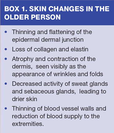
The epidermis, just 0.1mm thick, is very thin and is attached to the dermis at the dermo-epidermal junction. With increasing age, particularly after the age of 70, the epidermis gradually thins, leading to a flattened interface between the epidermis and the dermis, which reduces its resistance to shearing forces.3
The dermis is composed of connective tissue, blood vessels, lymphatics, macrophages, endothelial cells and fibroblasts. With increasing age, a reduction in collagen and elastin causes it to become more susceptible to external forces, such as friction and shear.
The subcutaneous layer consists of adipose and connective tissue that, as the skin loses elasticity and strength, becomes less resilient. As the fatty layer becomes thinner certain areas of the body, such as the face, neck and hands for example, will lack the cushioning produced by the fatty deposits and these areas can become more susceptible to skin tears.3
Skin changes in the older person are summarised in Box 1.
With age the skin can become very fragile and even a minor bump or knock can cause damage.4 The normal process of ageing thus increases the risk of skin tears. At the other end of the age spectrum, the very young with immature skin also have a higher risk.4
SKIN TEARS
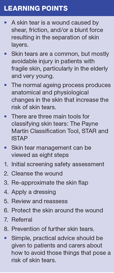
Skin tears have recently been defined as ‘… a wound caused by shear, friction, and/or a blunt force resulting in the separation of skin layers. A skin tear can be partial-thickness (separation of the epidermis from the dermis) or full-thickness (separation of both the epidermis and dermis from underlying structures).’5
Skin tears most frequently arise through trauma, and occur on the body’s extremities, including the lower limb, the arms and the dorsal aspect of the hands.6,7 When they occur on the front of the leg or on the shin bone they are classified as ‘pre-tibial lacerations’ and require assessment of the blood supply to the lower limb and consideration of the use of compression.4
The incidence rate of skin tears among patients over 65 years is estimated to be 15.5%,8 and 1.5 million skin tears occur in elderly residents of institutions in the USA annually.9 They are set to become one of the largest problems in wound care. It is therefore important to focus on prevention and creating a safe environment. Identifying and, where possible, removing factors that can cause skin tears can help to reduce prevalence, particularly in the elderly.
CLASSIFICATION SYSTEMS
There is no widely adopted, universally accepted classification system for the assessment of skin tears. There are, however, three main tools available:
- The original Payne and Martin classification tool, updated in 1993,10
- The Skin Tear Audit Research (STAR) Classification System,11 and
- The International Skin Tear Advisory Panel (ISTAP) Skin Tear Classification Consensus.2
The Payne and Martin tool10
Category 1: Skin tear without loss of tissue. These may be linear-type lesions with separation of the epidermis and dermis or flap-type where an epidermal flap covers the dermis to within 1mm of the wound margin.
Category II: The skin tear is subdivided into scant loss of tissue (25% or less) and moderate to large loss of tissue, with more than 25% loss of the epidermal flap.
Category III: The most severe type of skin tear with the loss of the entire epidermal skin flap. This may be caused by the initial trauma or necrotisation of the skin flap.
STAR classification11
The STAR classification includes consideration of the skin and flap colour.
Star category skin tears are classified as follows:
1a. A skin tear where the edges can be re-aligned to the normal anatomical position, without undue stretching. The skin or flap colour is not pale, dusky or darkened.
1b. A skin tear where the edges can be re-aligned to the normal anatomical position, without undue stretching. The skin or flap colour is pale, dusky or darkened.
2a. A skin tear where the edges cannot be re-aligned to the normal anatomical position, without undue stretching. The skin or flap colour is not pale, dusky or darkened.
2b. A skin tear where the edges cannot be re-aligned to the normal anatomical position, without undue stretching. The skin or flap colour is pale, dusky or darkened.
3. A skin tear where the skin flap is completely absent.
ISTAP2
ISTAP classifies skin tears into three types:
1. Type 1 – No skin loss. A Type 1 skin tear is a linear tear where the flap can be repositioned to cover the wound bed.
2. Type 2 – Partial flap loss. In a Type 2 skin tear, partial flap loss means that the skin flap cannot be repositioned to cover the wound bed.
3. Type 3 – Total flap loss. A Type 3 skin tear involves total flap loss that exposes the entire wound bed. Please note that deep Type 3 skin tears (i.e. total thickness wounds) will require specialist intervention, so this standard guidance does not apply.
ASSESSMENT AND CATEGORISATION
Wound assessment is necessary to establish the type of injury, with a focus on the prevention of further injury,1 and to determine:
- Location
- Dimensions (length, width and depth)
- Percentage of viable/non-viable tissue
- Degree of flap necrosis
- Presence of any haematoma
- Type and amount of exudate
- Integrity of surrounding skin.
Skin tears can be painful, with trauma affecting the superficial nerve endings both in and around the wound. The assessment and management of pain is essential,4 and pain measurement tools, such as the visual analogue scale (VAS) can be used to assess pain from ‘no pain’ to ‘worst possible pain’.12
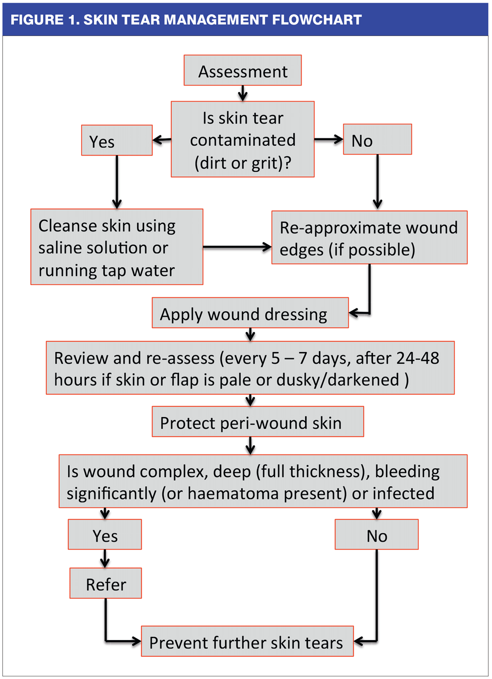
SKIN TEAR MANAGEMENT
Management of skin tears follows a number of steps, outlined below and shown in the algorithm (Figure 1).
1. Initial screening safety assessment
The initial screening should be about the patient’s overall safety.
2. Cleanse the wound
Use saline or running tap water to remove any dirt or grit and to control bleeding. Then gently pat dry the surrounding skin
3. Re-approximate the skin flap
- If the skin flap is viable, bring the edges together by gently easing the flap back into place using tweezers, a gloved finger, or a silicone strip. Record any approximation.
- For flaps that are difficult to align, consider using a moistened non-woven swab, applied for 5-10 minutes to rehydrate the area.
- Avoid using wound closure strips as they may cause traction and further trauma.
- Sutures and staples are not recommended due to the fragility of the skin.
- Apply a skin barrier product, as appropriate, to protect the surrounding skin.
4. Apply the dressing
A good wound management dressing should remain in place, prevent skin trauma on removal, and maintain an optimal moisture balance.13,14 In addition, it is essential that the dressing provides an environment that supports wound healing.
- If the flap cannot be approximated consider the use of technology lipido colloid (TLC) dressing to aid the formulation of new tissue within the wound bed.15
- After securing the flap, select a soft silicone or TLC dressing and apply without any tension. Choose a dressing appropriate for the wound condition and category of skin tear, ensuring a 2cm overlap around the wound.
- The wear time will be dependent on the type of dressing and amount of exudate. If possible the dressing should be left in place for up to 5 days to avoid disturbance of the skin flap. Mark the dressing with an arrow to indicate the direction in which it should be removed.
5. Review and reassess
- At each dressing change (approximately every 5-7 days), gently lift and remove the dressing, working away from the attached skin flap. Silicone-based adhesive removers can be used to avoid trauma to the surrounding skin.16
- When removing the dressing, observe and evaluate the wound and take care not to disrupt the skin flap
- Monitor for changes in the wound status and ensure maintenance of skin integrity. Where the skin or flap is pale and dusky/darkened, it is important to reassess within 24-48 hours as further breakdown may occur
- Monitor for any signs of infection and manage appropriately according to best practice guidelines.17
- Use digital photography where possible to document the wound.
- Treatment can be discontinued when complete epithelialisation occurs.
6. Protect the skin around the wound
- Protection of the skin is vital in maintaining its integrity. Keep the skin well hydrated by maintaining nutritional intake and fluid balance.
- Use pH friendly soap and pat dry
- Patients with dry skin on their arms and legs will benefit from the application of an appropriate pH friendly moisturising cream, applied twice a day.6,18
- Reduce moisture from incontinence or other sources
- Use caution when applying an adhesive tape directly on to the skin.
- Protect fragile skin by covering with tubular or roller bandages, long-sleeved clothing etc.
7. Referral
- Some complex skin tears or associated full thickness skin injury, significant bleeding or haematoma formation, may require surgical review and intervention to repair the injury.
- Caution should also be exercised if there is concern regarding blood clotting ability and the general blood supply.
- An inter-professional and collaborative approach to management is required to optimise healing outcomes for the individual.
8. Prevention of skin tears
Most skin tears occur during routine patient care activities. It is therefore important to bear this in mind and try to encourage the creation of a safe environment.
CREATING A SAFE ENVIRONMENT
Advice on the identification and removal of factors that cause skin tears can help to reduce their prevalence, particularly in the older person. An awareness of risk, by both patients and carers, should be encouraged and you can help them reduce the risk of skin tears with some simple, practical advice:
- Make sure there is adequate lighting and that light switches are easy to reach
- Avoid trip hazards, such as rugs and excessive furniture
- Ensure that small furniture (night table, chairs) in the immediate surroundings are positioned carefully to avoid unnecessary bumps or knocks
- Upholster the sharp borders of furniture or bed surroundings with padding and soft material
- When appropriate give carers advice about how to handle and move patients; e.g. never use sheets to move patients as this can contribute to damage by dragging on the skin. Local manual handling guidance should be followed.
- Reduce or eliminate pressure, shear and friction by using appropriate pressure relieving devices and positioning techniques
- Always wear appropriate footwear and clothing to reduce the risk of injury
- Wearing long socks can protect the pre tibial area
- Avoid acrylic adhesive dressings where there is a risk of a skin tear
- Remove dressings slowly to avoid tearing the skin.
SUMMARY
It is important that you have an understanding of the effects of ageing on the skin. This knowledge will enable you to advise patients and carers appropriately about the measures they can take to reduce the risk of skin tears.
Skin tears are common and, for those patients who sustain them, assessment, categorisation, management skills and documentation are important for effective care. High standards of appropriate care can reduce the need for hospital appointments and admissions. For our patients, effective care can help them avoid the pain and potential complications of this injury, including chronic wounds, leg ulceration and cellulitis.
REFERENCES
1. Stephen-Haynes J, Carville K. Skin Tears Made Easy. Wounds International 2011; 2(4):1–6
2. LeBlanc K, Baranoski S, Christensen D, et al. International Skin Tear Advisory Panel: A tool kit to aid in the prevention, assessment, and treatment of skin tears using a simplified classification system. Advances in Skin and Wound Care 2013;26(10):459–76
3. Sibbald RG, Krasner DL, Lutz JB, et al. The SCALE Expert Panel: Skin Changes At Life’s End. Final Consensus Document. October 1, 2009.
4. Beldon P. Classifying and managing pretibial lacerations in older people. British Journal of Nursing 2008;17(11):Tissue Viability Supp S4 -S18
5. LeBlanc, K. Baranoski, S. Skin Tears: State of the Science. Consensus Statements for the Prevention, Prediction, Assessment, and Treatment of Skin Tears. Advances in Skin and Wound Care 2011; 24(9):2-15
6. Carville K, Leslie G, Osseiran-Moisson R, et al. The effectiveness of a twice-daily skin-moisturising regimen for reducing the incidence of skin tears. International Wound Journal 2014;11:446–53
7. Baranoski, S. How to prevent and manage skin tears. Advances in Skin and Wound Care 2003;16(5):268-70
8. Konya C, Sanada H, Sugama J et al. Skin injuries caused by medical adhesive tape in older people and associated factors. Journal of Clinical Nursing 2010;19: 1236–42
9. Baranoski S. Meeting the challenge of skin tears. Advances in Skin and Wound Care 2005;18(2):74-75
10. Payne RL. Martin ML. Defining and classifying skin tears: need for a common language. A critique and revision of the Payne-martin classification system for skin tears. Ostomy Wound Management. 1993;39 (5):16-26
11. Carville K, Lewin G, Newall N, et al. STAR: A consensus for skin tear classification. Primary Intention 2007;15(1):18-28
12. Mudge E, Orsted H. Wound infection and pain management made easy. Wounds International May 2010;1(3):1-6 www.woundsinternational.com/media/issues/290/files/content_8902.pdf
13. Okan D, Woo K, Ayello EA et al. The Role of Moisture Balance in Wound Healing. Advances in Skin and Wound Care. 2007; 20(1):39-53.
14. World Union of Wound Healing Societies (WUWHS). Principles of best practice: Wound exudate and the role of dressings. A consensus document. MEP Ltd. London, 2007.
15. White R, Cowan T, Glover D. Evidence-based dressing selection. Journal of Wound Care http://dx.doi.org/10.12968/jowc.2011.20.Sup1.4
16. Beldon P. Skin Trauma. In: White R, Harding K (eds) Trauma and Pain in Wound Care. Wounds UK, Aberdeen 2006
17. Wounds UK. Best Practice statement: the use of topical antimicrobial agents in wound management. Wounds UK. London; 2013 (3rd Edition)
18. Wounds UK. Best Practice Statement. Care of the Older Person’s Skin. Wounds UK; London 2012

Related articles
View all Articles