Healing wounds: where can it go wrong?
ELIZABETH MERLIN-MANTON
ELIZABETH MERLIN-MANTON
(RGN; MSc; TVN)
Tissue Viability Nurse;
Manchester Foundation Trust
The length of time it takes a wound to heal can be influenced by many factors, including the initial treatment plan, differential diagnosis, selection of appropriate treatment and the expediency of specialist referral when needed
From the entire UK population of 63.7 million people, mid 2013, it was estimated that there were 2.2 million patients with a wound.1
The longevity of any wound type can be influenced not only by underlying comorbidities but by the initial treatment plan, speed of differential diagnosis, selection of appropriate treatment and the expediency of specialist referral.
The annual prevalence of wound types, (e.g. burns, surgical, traumatic, ulcers) has been analysed across the UK population with an average of 61% of all wounds achieving full healing within a 12 month period. However, only 43% of chronic wounds healed within this timeframe contrasting with 79% of acute wounds.1 Within this analysis unspecified leg ulceration was identified as the highest single wound category at 19% and collectively lower limb ulcers, encompassing venous, arterial, mixed aetiological, unspecified leg ulcers and diabetic foot ulcers, represented 42% of all wound types, equalling 900,000 wounds.1
For lower limb ulcerations, it is evident that many take more than 12 months to heal,2,3 and some result in devastating effects. Practice nurses and other generalist community clinicians have the ability to play a vital role in improving patient outcomes by gaining an enhanced awareness, enabling earlier detection of the differential diagnosis, resulting in prompter, pertinent, intervention.
The ideal wound healing process consists of:
- Haemostasis where platelets clot to minimise blood loss – lasting for a few hours;
- Inflammatory response to aid removal of wound debris by autolysis – lasting up to 3 days;
- Proliferation which includes angiogenesis, epithelialisation and granulation – lasting up to 21 days;
- Maturation – cell reorganisation and development of scar tensile strength – for up to 2 years.
During wound healing, these phases overlap and although estimated time frames are given, the evidence has demonstrated many wounds will not advance successfully and may become uncontrolled. Therefore, all clinicians involved in wound care require the ability to correctly identify the current phase of wound healing to enable the correct choice of treatment and specialist referral if required.
PATHWAYS
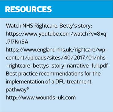
National expert guidance or local specialist trust recommendation, in the form of treatment algorithms, clinical pathways and referral pathways are often used to provide comprehensive, effective care,4 and have been demonstrated to enhance patient outcomes while simultaneously having a positive economic impact.5 For the generalist wound care nurse, this guidance can be invaluable and NHS England has provided clinicians with their RightCare scenario of Betty’s Story,8 which considers both an optimal and suboptimal patient pathway.
RAG rating (Red, Amber, Green) is frequently used by the specialist during the creation of a clinical pathway as a point of focus to guide a clinician’s consideration to the present wound state and required action.5,6 The RAG system can be used to direct either a suggested change in the treatment provided or a recommendation that a specialist referral is made.
LEG ULCERS
Early recognition of a wound’s aetiology allows the most suitable action to be taken and although many healthcare clinicians involved in wound care recognise that the loss of skin below the knee, on the leg or foot, as an ulcer, the differential diagnosis is often undetected.
Nonetheless, due to the underlying comorbidities these wounds are deemed as chronic if they take more than 2 weeks to heal.7 Any wound is at risk of becoming chronic from initial manifestation and a recurring wound could be considered at risk from onset.
Venous valves, that are designed to prevent venous return, can become damaged or incompetent due to predisposing risk factors such as occupation, past medical history or existing comorbidities, thus resulting in venous disease, venous hypertension and chronic venous insufficiency (CVI). There are various different phases of CVI commonly confirmed with visible characteristic signs that are typically overlooked. These visible signs include:
- Oedema that worsens during the day
- Varicose eczema
- Spider veins (telangiectasia)
- Varicose veins, and
- Other skin changes such as haemosiderin staining.
During a routine health review, if symptoms such as pruritus, or the visible signs are noted, compression hosiery should be a consideration to avoid further deterioration. With an open wound present, the optimal pathway, as in the NHS Right Care pathway, Betty’s story, recommends compression hosiery to be prescribed if a risk assessment has ruled out arterial disease, with a referral for a full, vascular assessment at a specialist clinic.8,9
As venous leg ulcers are slow to progress, repeatedly patients relay the story of a lengthy, yet slow deterioration and often an active ulcer is present by the time the patient presents themselves at their GP surgery, indicating the final phase of CVI.
Differentiating between ulcer types is possible with the presence or lack of the distinctive signs. A venous ulcer is usually positioned above the malleolus and below the knee, while an arterial ulcer is usually on the extremities of the lower limb. Arterial ulcers have clearly defined edges and the onset is rapid: the skin has a shiny appearance and is cool to touch, and compression is contraindicated.
Unsurprisingly, mixed aetiology ulcers occur whereby a combined range of signs and symptoms are apparent.
DIABETIC FOOT ULCERATION
Around 10% of people with diabetes will suffer a diabetic foot ulcer (DFU) at some point in their lives. DFU is defined as a localised injury to the skin and/or underlying tissue, below the ankle, in a person with diabetes.10 Accurate diagnosis and appropriate treatment is essential for achieving the best outcome for the patient,7 and can reduce a DFU’s longevity, while undoubtedly impacting on the patient’s quality of life.11,12 It is crucial that DFU aetiology is ascertained to establish appropriate management, therefore referral to a specialist clinician is essential. NICE guidance (NG19) recommends referral of a person with diabetic foot problems within 24 hours, therefore being aware of the local referral policies is beneficial to all practitioners, especially as they vary and include either a foot protection team or a Multi-Disciplinary Team. In the recent National Diabetes Footcare Audit (NDFA) report,13 it was highlighted that late referrals lead to a poorer prognosis in terms of infection and amputation, emphasising the importance of prompt referrals.
ESSENTIAL SKIN CARE
Although patients with wounds can be any age, it must be noted that the aged skin can present challenges, such as a decline in the epidermal and dermal thickness. It is vital that even while waiting for specialist involvement or further assessments, such as a Doppler assessment to ascertain the ankle brachial pressure index (ABPI), the essential wound and skin care is implemented. Protection is one of the skin’s main functions but a patient can become vulnerable if this protective function is weakened and the skin becomes too dry. Therefore, it is essential that the skin, particularly on the lower limbs, is cleansed – usually with a soap substitute – and hydrated by means of a topical emollient.
WOUND ASSESSMENT
In addition, the wound requires a thorough assessment to identify any factors that may interrupt healing. This requires documenting in the patient’s notes or on the wound assessment charts available to the clinician. The wound should be measured at each dressing change and if local policies allow, with patient consent, a wound image should be considered.
Documenting the tissue type percentage facilitates an indication of progress, deterioration or whether the wound remains unchanged. Generally the tissue type classifications used are: necrosis, slough, infection, granulation or epithelial tissue, although there are many other useful descriptors.
During the inflammatory phase of wound healing matrix metalloproteinases (MMP) are present. MMP activity is controlled by tissue inhibitors and the equilibrium between these dictate wound progression. It is recognised that high MMP levels correlate with delayed wound healing and there can be up to 65 times more MMPs present in chronic wounds than acute wounds.14
Exudate is mostly produced during the inflammatory or proliferative phases and exudate levels can provide an indication of wound status. Wound type, location and size also influence these levels.15 However, if high levels of exudate persist it is likely to be caused by the underlying aetiology, such as an imbalanced bacterial burden or oedema. Detailing whether the wound is dry, moist, wet, saturated or leaking is an essential part of the wound assessment as exudate levels can influence wound progression.16
The wound infection continuum provides guidance as to the signs and symptoms that correlate with each stage.17 These stages are contamination, colonisation, local infection, spreading infection and systemic infection. Contamination and colonisation require observation but not direct action, but for local infection a topical antimicrobial is recommended, and for spreading or systemic infection both a systemic and a topical antimicrobial are required.
Additional important sections of a wound assessment are the wound edges and the surrounding skin. Observing the status of the wound edge supports the understanding as to whether epithelial advancement will be delayed. Rolled, dry or macerated wound edges have the potential to delay wound healing. In addition to dry or macerated peri-wound areas, there are other considerations such as varicose eczema, fragile elderly skin and excoriation, all of which require effective management to prevent interruption of the wound healing process.
EVIDENCE BASED PRACTICE
Evidence based practice informs clinical decisions and clinical research is valuable in supporting it,6,18 but robust evidence is limited within the wound care arena. Therefore, by utilising validated clinical pathways, immediate evidence based treatments could be used from onset, thus minimising a further risk of delayed wound healing, as a concurrent referral to the specialist clinician has been made.
SUMMARY
Caring for patients with wounds is a complex process and living with a wound significantly impacts a patient’s quality of life. Wound healing and earlier referrals must improve to avoid the increasing financial burden to the NHS,19 and the importance of the general practice nurse’s role should not be underestimated in relation to reducing the time to healing through earlier intervention and specialist referral.
REFERENCES
1. Guest JF, Ayoub N, McIlwraith T, et al. Health economic burden that wounds impose on the National Health Service in the UK. BMJ Open 2015. https://bmjopen.bmj.com/content/5/12/e009283
2. Meaume S, Dompmartin A, Lok C, et al. Quality of life in patients with leg ulcers: Results from CHALLENGE, a double-blind randomised controlled trial. Journal of Wound Care 2017; 26(7):368-379. 10.12968/jowc.2017.26.7.368
3. Green J, Jester R, McKinley R, Pooler A. Patient perspectives of their leg ulcer journey. J Wound Care 2013;22(2):58-66. 10.12968/jowc.2013.22.2.58
4. Gardner S. Using treatment pathways to improve healing of venous leg ulceration. Wounds UK 2013;9(1):67–75
5. Mullings J, Merlin-Manton E. Improving patient outcome through the implementation of a person-centred leg ulcer pathway. J Wound Care 2018;27(6):378 https://doi.org/10.12968/jowc.2018.27.6.378
6. Best practice recommendations for the implementation of a DFU treatment pathway. London: Wounds UK, 2018. https://www.wounds-uk.com/resources/all/20/date/desc
7. NICE Clinical Knowledge Summaries; Leg ulcer – venous. February 2016
8. NHS England Right Care: Betty’s story. https://www.england.nhs.uk/rightcare/wp-content/uploads/
sites/40/2017/01/nhs-rightcare-bettys-story-narrative-full.pdf
9. Wounds UK. Best Practice Statement: Compression hosiery (2nd edn), 2015. https://www.wounds-uk.com/resources/all/0/
date/desc/keyword/Compression%20hosiery
10. NICE NG19: Diabetic foot problems: prevention and management, 2015 (updated January 2016). https://www.nice.org.uk/guidance/NG19
11. Oyibo S, Jude E, Tarawneh I, et al. The effects of ulcer size and site, patient’s age, sex and type and duration of diabetes on the outcome of diabetic foot ulcers. Diabetic Medicine 2001;18:133–8
12. Merlin-Manton E, Mitchell L, Whalley A. The importance of limiting diabetic foot ulcer chronicity The Diabetic Foot Journal 2016;19:156–61
13. NHS Digital. National Diabetes Foor Care Audit. March 2018 https://digital.nhs.uk/data-and-information/publications/statistical/national-diabetes-footcare-audit/national-diabetes-foot-care-audit-2014-2017
14. Lazaro JL, Izzo V, Davies AH, et al. Elevated levels of metalloproteinases and chronic wound healing: an updated review of clinical evidence. J Wound Care 2016;25(5):277-87
15. Cutting KF. Wound exudate: composition and functions. Br J Community Nursing 2003;8(Suppl 3):11577
16. Romanelli M, Dowsett C, Doughty D, et al. Position Document. Advances in wound care: the triangle of wound assessment. Wounds International 2016; http://www.woundsinternational.com/wuwhs/view/position-document-advances-in-wound-care-the-triangle-of-wound-assessment
17. Swanson T, Angel D, Sussman G, et al. Wound infection in clinical practice. International Wound Infection Institute. Wounds International 2016. http://www.woundsinternational.com/consensus-documents/view/iwii-wound-infection-in-clinical-practice
18. Greenhalgh T. How to read a paper: The basics of evidence-based medicine. (5th ed). Oxford: Blackwell Publishing; 2014
19. Guest JF, Vowden K, Vowden P. The health economic burden that acute and chronic wounds impose on an average clinical commissioning group/ health board in the UK. J Wound Care 2017;26(6):292-303
Related articles
View all Articles