QOF perspective: Management and prevention of osteoporosis
Anne Sutcliffe B.Sc.(Hons) RGN, DN
Anne Sutcliffe B.Sc.(Hons) RGN, DN
Healthcare and Education Officer Paget's Association
While osteoporosis is often perceived to be a 'female' disease, it affects men too. With osteoporosis recently added to the Quality and Outcomes Framework, it is a good time to think harder about this debilitating and life threatening condition.
It is estimated that osteoporosis occurs in approximately 3 million people in the UK and results in more than 230,000 fractures per annum with the commonest ones occurring at the hip, vertebral body and forearm.
Approximately 75,000 hip fractures occur annually in the UK1 with the average age of incidence being 84 and 83 years in men and women respectively.2 There is an estimated 30% probability of death during the first year after fracture and approximately half the survivors will suffer from long term disability and loss of independence.3
Accurately quantifying the incidence and prevalence of vertebral fractures is difficult, as many patients with back pain do not seek medical attention and there has also been a lack of a universally recognised definition of vertebral deformity from X-rays. In the UK it has been estimated that the lifetime risk of a vertebral fracture in a 50-year-old woman is 3.1% whereas the corresponding figure for a 50-year-old man is 1.2%.4
Effective treatment options are available for both primary and secondary prevention of fragility fractures in postmenopausal women. These have been subject to several lengthy appraisals by NICE in recent years, with the recommendation that alendronic acid (a bisphosphonate) should be given following fracture and also used in primary prevention when there are significant risk factors and evidence of reduced bone density. Other therapies may be considered if an individual cannot tolerate alendronate.5,6
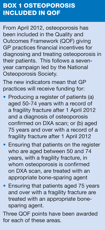
Secondary prevention of osteoporotic fractures has been included in the Quality and Outcomes Framework (QOF) since April 2012, but there is additional scope for practice nurses to consider incorporating osteoporosis risk assessment into other areas of chronic disease management.
Case study
While attending for review of type 2 diabetes, Mrs D, who is 63 years old, expresses concern about her risk of osteoporosis. Further questioning elicits the following reasons why she is worried.
- Type 2 diabetes for 2 years, currently controlled by diet and no present complications.
- Lower lumbar back pain for several years.
- Osteoarthritis in lower spine and both knees (confirmed radiologically).
- Colles' fracture aged 40 years (following a fall from a bicycle).
- Her twin sister has recently had a DXA scan following a Colles' fracture. This showed that she had reduced bone density in her spine and she has subsequently started treatment with alendronate.
Mrs D wants to know if she should have a bone density scan and whether her risk of osteoporosis is high. She is also interested to know if she needs to change her diet, consider additional calcium supplements and whether any type of exercise would help to prevent osteoporosis.
Using the information you have been given:
1. Does Mrs D present with any risk factors that would increase the likelihood of osteoporosis and fractures?
2. Are you aware of any assessment tool that may guide your management?
3. Would you suggest any further investigations?
4. What lifestyle advice would you offer?
Skeletal integrity and fracture risk are influenced by numerous agents including genetic, hormonal, endocrine and lifestyle factors. The importance of these factors varies, with some acting independently and others dependently with different relevance at different ages. Risk factor assessment, with or without bone density measurements, may help to guide you in assessing future fracture risk and the need for therapeutic intervention.
Diabetes
Insulin acts on receptors on bone cells to stimulate amino acid uptake and collagen synthesis hence acting as a skeletal growth factor. Insulin-dependent diabetics have been shown to have modest reductions in bone mineral density (BMD) at the spine and appendicular skeleton when compared with age matched controls. Although women with type 2 diabetes do not necessarily have a reduced bone density there does appear to be an increased risk of fracture. This could be associated with visual impairment and peripheral neuropathy increasing the risk of falls.7 Currently it is not recommended that women with type 2 diabetes should have bone density measurements performed routinely.
Back pain
The major clinical consequences of vertebral fractures are back pain, loss of height and kyphosis, all of which can lead to significant morbidity. Pain caused by vertebral fracture is usually associated with intense localised tenderness accompanied by paravertebral muscle spasm. This severe pain usually decreases in severity over several weeks or months but in a proportion of patients, persistent pain experienced while standing and bending may continue six months after fracture. As chronic low back pain can also be associated with a number of other conditions it is important to examine the back and question the patient about pain intensity, timing, associated stiffness and problems with mobility. While Mrs D's back pain could be caused by a vertebral fracture a previous spine X-ray has shown osteoarthritis, which is a more likely reason for her chronic pain.
Osteoarthritis
Osteoarthritis is not associated with osteoporosis. However, there is a twofold increase in osteoporosis in both male and female populations with rheumatoid arthritis compared with healthy controls, with bone loss being more marked in male than female patients. Bone loss is associated with osteoclastic activity due to the inflammatory disease process and may also be aggravated by the use of corticosteroids.
Colles' Fracture
It has long been apparent that those who have already sustained one fracture appear to be at a greater risk of subsequent fractures. Following an initial Colles' fracture approximately 20% of women will sustain a subsequent fracture within 1 year, rising to 60% at five years.8 However, this risk is greater if the initial fracture occurs after the age of 50 years and is not associated with trauma. As Mrs D's fracture occurred when she was 40 years, was trauma related, and she has had no subsequent fracture history it is unlikely that her future fracture risk will be any greater than normal for her age.
Family history
Osteoporosis is a disease with a strong heredity component and twin and family studies have shown that genetic factors contribute to osteoporosis by influencing bone density and other determinants of fracture risk, such as skeletal architecture and bone turnover. Many twin studies have demonstrated that females are likely to have a similar bone density when peak bone mass has been achieved around the early 20s. However, those studies that have considered postmenopausal bone density in twins have shown that the genetic component is somewhat lower. A parental history of hip fracture is associated with a doubling of the risk of sustaining a hip fracture in men and women. A large study of male and female twins from Finland has directly examined the heritability of fracture. This showed that the genetic component of fracture was relatively small at <30% and appeared to be smaller in women than men.9 Although genetic factors are important, it is also important to consider that fracture is also influenced by a number of other factors including muscle function, body size and shape, incidence of falls and other additional environmental factors.
MANAGEMENT
Although Mrs D may have been concerned about osteoporosis the points covered would suggest that her risk of osteoporosis is no higher than normal for her age and at this point in time she merely requires reassurance. There is no reason to perform a bone density scan or any other investigation and no need to consider treatment for reduced bone density.
Should further reassurance be required it would be worth considering the use of FRAXTM WHO Fracture Risk Assessment Tool, accessed at http://www.shef.ac.uk/FRAX
Use of this assessment tool provides an easy way of entering information about an individual's age, gender and risk factors for future fracture. FRAX is then able to calculate a figure indicating a 10-year fracture probability of both hip and other major osteoporotic fracture. The advantage of this specific tool is that it produces an accurate numerical value, presented in percentage terms that can be easily explained to the patient.
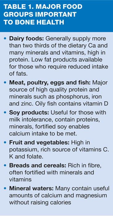
As part of her diabetes management Mrs D should have been given relevant dietary advice but it would be worth checking that she is eating a well balanced diet that will also optimise bone health. She needs at least 700mgs of calcium daily and a variety of foodstuffs to provide protein, vitamins and minerals to maintain skeletal integrity (Table 1).
Vitamin D is required to aid calcium absorption and also contributes to muscle strength, which in turn influences the propensity to fall. There are a few naturally occurring food sources of vitamin D but these are generally high in fat. Vitamin D synthesis in the skin from exposure to the sun is the primary determinant of the active form of this vitamin. It is therefore important to encourage Mrs D to go outdoors regularly in the summer months - approximately 20 minutes on a daily basis should be sufficient to maintain adequate supplies of vitamin D.
Provided Mrs D is eating a well balanced diet with adequate calcium and is able to go outdoors regularly and expose her face and arms to the sun she is unlikely to require extra calcium and vitamin D supplementation.
Activity can help to maintain bone density and also has positive effects on muscle mass, balance, risk of falling, morbidity, confidence and quality of life. To achieve maximal benefit, regular physical activity (weight bearing, endurance and strength training) must target vulnerable bone sites and be progressive in intensity. Ideally, activities should be sustained over time as bone loss will occur once exercise is stopped.
Advancing age itself increases the risk of osteoporotic fracture and should Mrs D sustain an atraumatic fracture, experience falls, become immobile and housebound, further assessment would be required. In addition, poor control of her diabetes or related complications could adversely affect bone health and this should be considered as part of her diabetic management plan.
CONCLUSION
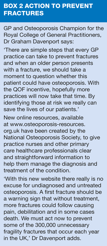
Given the long-term morbidity and increased mortality associated with osteoporotic fractures, the possibility of osteoporosis is something that practice nurses should consider in patients with risk factors, and as part of their management of other long-term conditions, including diabetes. Resources are now available to make it easier to calculate risk, without resorting to DXA scanning where this is less likely to be necessary.
REFERENCES
1. British Orthopaedic Association (BOA) and British Geriatrics Society (BGS). The Care of Patients with Fragility Fracture: BOA, 2007
http://www.fractures.com/pdf/BOA-BGS-Blue-Book.pdf
2. National Hip Fracture Database. The National Hip Fracture Database Report (Extended Version). 2010 http://www.nhfd.co.uk/003/hipfractureR.nsf/NHFD_National_Report_Extended_2010.pdf
3. Parker M, Johansen A. Hip fracture. British Medical Journal 2006, 333(7557):27-30
4. Van Staa TP, Dennison EM, Leufkens HG et al. Epidemiology of fractures in England and Wales. Bone 2001; 29: 517-22.
5. NICE TAG 160. Alendronate, etidronate, risedronate, raloxifene and strontium ranelate for the primary prevention of osteoporotic fragility fractures in postmenopausal women. 2011
6. NICE TAG 161. Alendronate, etidronate, risedronate, raloxifene, strontium ranelate and teriparatide for the secondary prevention of osteoporotic fragility fractures in postmenopausal women.2011
7. Brown SA, Sharpless JL. Osteoporosis. An under-appreciated complication of diabetes. Clinical Diabetes 2004; 22 (1): 10-20.
8. Cuddihy MT, Gabriel SE, Crowson CS et al. Forearm fracture as predictor of subsequent osteoporotic fracture. Osteoporos Int 1999; 9: 469-75
9. Kannus P, Palvanen M, Kaprio J et al. Genetic factors and osteoporotic fracture in elderly people; prospective 25 year follow up of a nationwide cohort of elderly Finnish twins. BMJ 1999; 319: 1334-7.
Related articles
View all Articles


