Tropical infectious diseases
Dr Hector Maxwell-Scott
Dr Hector Maxwell-Scott
MSc DTMH MRCP FRCPath
Imported Fever Service and the Hospital for Tropical Diseases
Many infections acquired by travellers when abroad can present with similar symptoms, most notably fever, and without specialist investigation it can be difficult to determine the most appropriate approach to management
Over the last two decades international travel has become increasingly common, the Department for Transport estimates that in the United Kingdom (UK) over 240 million international flights left the UK in 2017, up by 50 million compared with 2007.1 This increase in travel puts greater numbers of people at risk of tropical infectious diseases and therefore healthcare professionals must be vigilant to best manage imported infections.
IMPORTED FEVER SERVICE
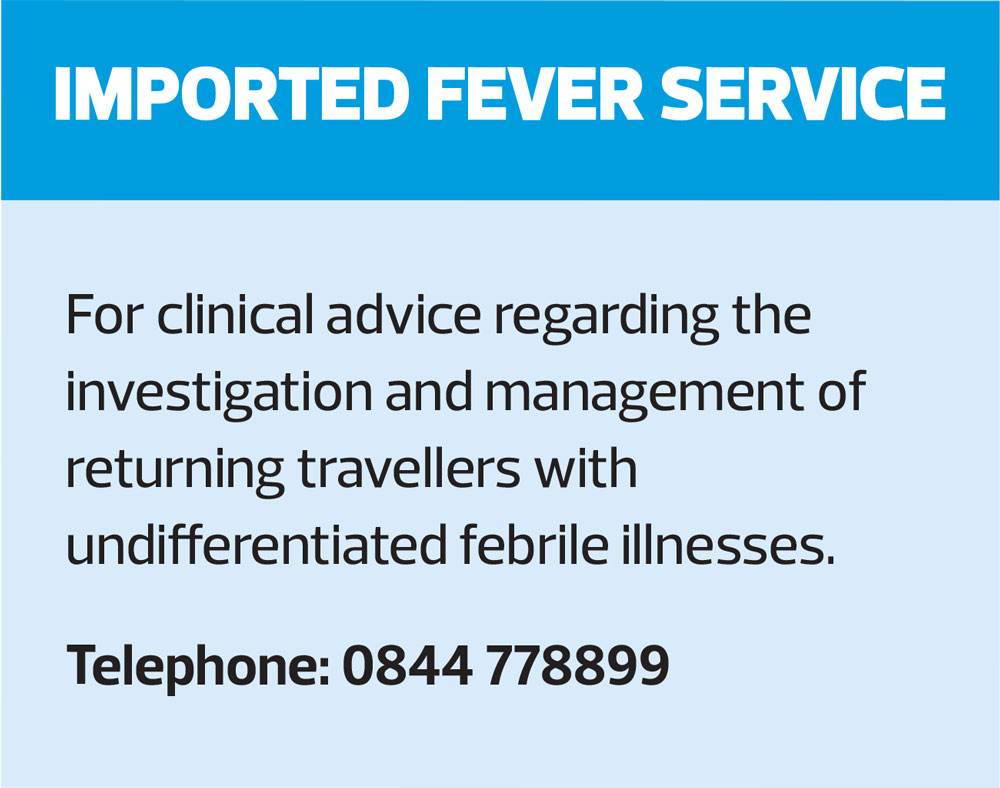
The Imported Fever Service (IFS) is a diagnostic and clinical advice service which is run from Public Health England’s (PHE) Rare and Imported Pathogens Laboratory (RIPL) in partnership with the Hospital for Tropical Diseases, London and the Tropical and Infectious Diseases Unit, Liverpool. It has been in operation since 2012 and provides advice to infection specialists across the UK regarding all aspects of the investigation and management of patients with undiagnosed imported febrile illnesses.
As many imported infections present with similar symptoms, testing is performed using geographic panels which cover the likely pathogens a patient may have encountered in a given area, along with additional testing added based on the exposure history and symptomatology. This unique strategy ensures that where possible, a diagnosis is made promptly allowing for appropriate management to be instigated in a timely fashion.
Clinical cases should be discussed with a local infection specialist (microbiologist, virologist or infectious diseases physician) in the first instance. If required an IFS clinician is available 24 hours a day, seven days a week via the on-call telephone number to offer advice on diagnosis, infection control and clinical management for imported fevers. In 2017 a total of 741 calls were received by the IFS, relating to 280 clinical cases.
THE APPROACH TO THE RETURNING TRAVELLER
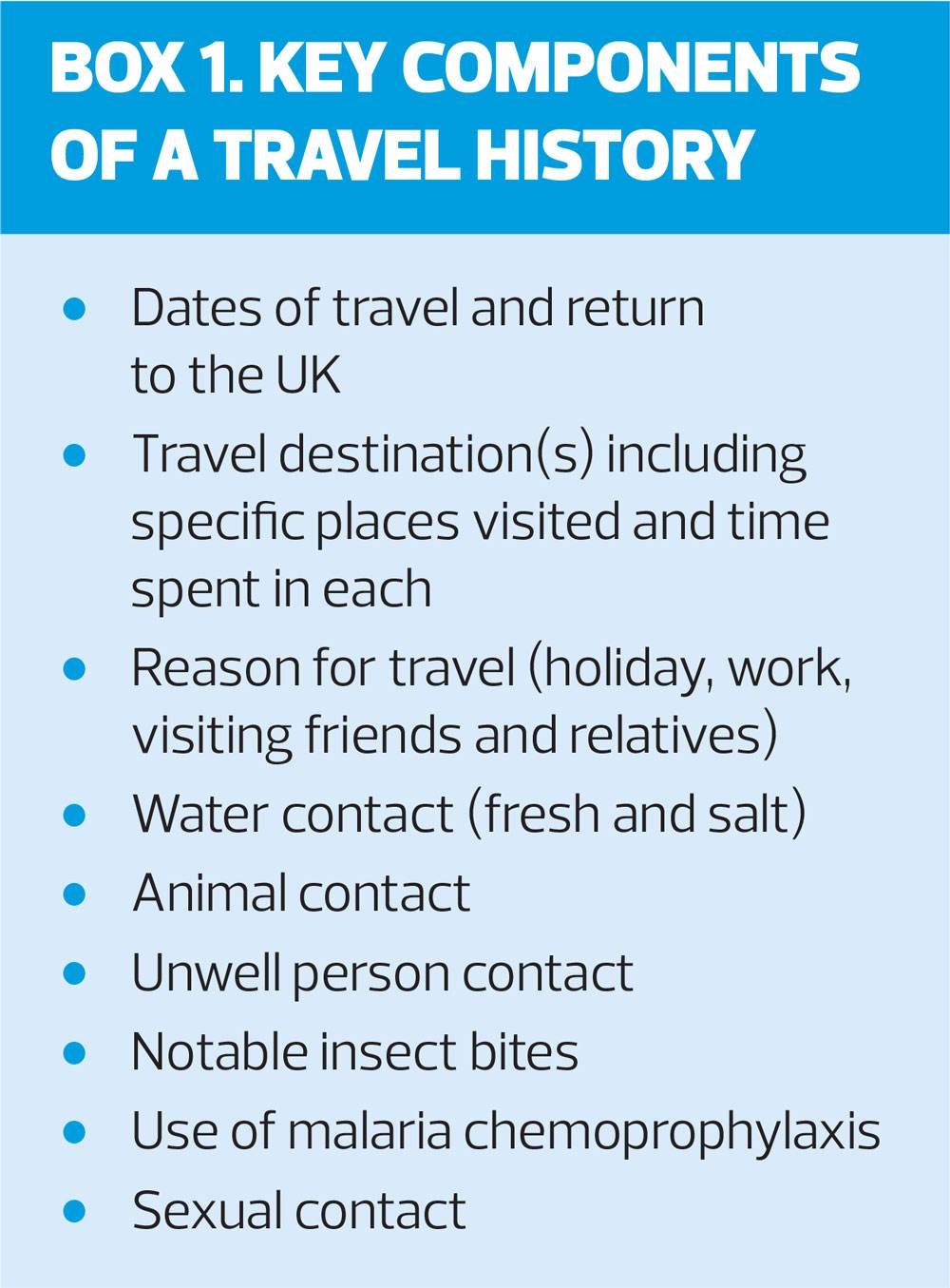
The ability to elicit a complete and accurate travel history is vital for healthcare professionals managing patients with possible imported infections (Box 1). The time between return to the UK and symptom onset is fundamental when considering possible infections, as an example most viral infections will present within 14-21 days of travel while parasitic infections such as malaria can present much later. Activities undertaken and locations visited while abroad also have a significant impact on the possible causes of fever in the returning traveller; fresh water exposure is a risk factor for leptospirosis and schistosomiasis, and visiting bat caves may increase the risk of contracting histoplasmosis, rabies and Ebola virus disease. Bites from insects and other arthropods as well as animal contact may also give clues about the possible causes of fever in a returning traveller.
In addition to considering tropical diseases, it is also important to remember that patients who have travelled recently may have acquired an infection in the UK and screening for routine pathogens should also be performed. Furthermore, the patient presenting with fever may not be suffering from an infection at all; lateral thinking is key to achieving a diagnosis.
IMPORTED INFECTIONS
Malaria
Malaria is an infection caused by single-celled parasites of the genus Plasmodium which are spread to humans via the bite of an infected female Anopheles mosquito. P. falciparum is the most common species with the highest associated mortality.2 Throughout history malaria has killed more humans than any other infection and even today it cases a significant burden of disease: the World Health Organization (WHO) estimates that in 2018 there were 219 million cases of malaria globally, with 435,000 deaths.3
Infection is characterised by fever, myalgia and rigors. Presentation may be entirely non-specific, so it is not possible to diagnose malaria based on symptoms alone; testing is therefore recommended for all patients with fever who have returned from a malaria risk area within the last six months (and indeed may be indicated when the date of travel was even earlier). Blood film analysis is the mainstay of diagnosis with testing complemented by rapid diagnostic assays for parasite antigen detection.
Treatment for malaria should always be discussed with an infection specialist and admission to hospital is recommended for the majority of patients with infection caused by P. falciparum. In addition to treatment for the acute infection, treatment is also required for eradication of dormant parasite forms in infection caused by P. vivax and P. ovale spp.
The viral haemorrhagic fevers
The viral haemorrhagic fevers (VHF) are a group of viral infections, examples of which include Ebola virus disease, Lassa fever and Yellow Fever. All present initially as an undifferentiated febrile illness with bleeding a late feature if present at all. Transmission to humans usually occurs via close contact with the reservoir host, an arthropod vector or via contact with an infected person.
Imported cases of viral haemorrhagic fever are exceptionally rare in the UK, however a careful risk assessment is required for patients presenting with fever within 21 days of return from a risk area. PHE and the Advisory Committee on Dangerous Pathogens (ACDP) provide a risk assessment algorithm4 for such patients and healthcare professionals should have a low threshold for discussing cases with their local Infection specialist if there is any doubt about the risk of a VHF.
Where necessary cases should be discussed with the IFS and urgent testing for VHF can be performed at RIPL 24 hours a day, seven days a week. Patients with confirmed VHF infection will be transferred to a UK centre specialising in the management of patients with high consequence infectious diseases.
Arboviruses
Arbovirus is a broad term that includes all viruses transmitted via an arthropod vector. Common examples imported in to the UK are dengue, chikungunya, Zika and Toscana virus. Dengue is found through much of the tropics and is the most commonly diagnosed arbovirus in the UK. Typically, infection causes an acute onset febrile illness with associated retroorbital headache and a widespread macular rash. Blood tests may show thrombocytopaenia and mild-to-moderate transaminitis. Chikungunya causes a similar acute illness, often without thrombocytopaenia, but may result in a prolonged arthralgia. Similarly, Zika virus infection can present with an undifferentiated illness with fever, although most cases are asymptomatic. Rarely, Zika virus infection may precipitate Guillain-Barre syndrome and infection during pregnancy has been associated with congenital microcephaly. All three viruses are transmitted via the bite of the female Aedes mosquito which feeds during the day and is frequently found in urban settings.5
Toscana virus is transmitted via the bite of the Phlebotomine sand fly and is found throughout much of southern Europe. While the majority of infections are likely asymptomatic, Toscana virus may cause meningitis, indeed it is one of the most common cause of a viral meningitis in southern Italy during the summer months. Infection should therefore be considered in patients return from the Mediterranean basin with meningitis.
Helminths
Helminths (worms) are a broad group of parasites causing infection in humans. Strongyloidiasis, caused by Strongyloides stercoralis, is the most common imported helminth infection in the UK. The larval stage is found in grass and soil in tropical and subtropical regions and is able to penetrate intact skin to enter the bloodstream. Following a migratory stage the adult worm settles in the wall of the gastrointestinal tract where it lays eggs that either pass in to the environment with stool or hatch to release larvae within the bowel, and which may then re-enter the host. Infection is therefore able to persist for decades post-exposure. Acute infection may be asymptomatic or may be associated with a rapidly moving urticarial rash (larva currens). While the adult worms do not usually cause symptoms, fever and wheeze may result from the migratory stage. Blood testing may reveal eosinophilia and all patients with eosinophilia and a relevant travel history should be screened for strongyloidiasis regardless of when they travelled.
Schistosomiasis is an infection caused by one of several species of fluke belonging to the genus Schistosoma. Free-living forms are found in bodies of fresh water and are able to penetrate unbroken skin to enter the bloodstream. Acute infection may be asymptomatic or present with fever, widespread urticarial rash and wheeze (Katayama fever). Symptoms resolve as the adult worms settle in the vascular plexus supplying the gastrointestinal tract or bladder (depending upon the species). Blood tests typically show an eosinophilia with eggs of the parasite being detectable in stool or urine. High burden chronic infection may result in portal hypertension or bladder cancer, but these complications are very uncommon in travellers.6
Cutaneous larva migrans is an infection caused by non-human hookworm. The larvae are able to penetrate skin but not enter the bloodstream. Infection results in a characteristic pruritic, serpiginous rash which moves over a course of hours to days as the larvae migrate through the subcutaneous tissues. Infection is not serious but can be distressing for the patient. Diagnosis is made by recognition of the characteristic rash and treatment is highly effective.
Sleeping sickness
Human African Trypanosomiasis or sleeping sickness, is caused by two subspecies of the single celled parasite Trypanosoma brucei. Infection is transmitted by the tsetse fly, which has a painful bite that is often recalled by the patient. Two subtypes exist, one in West Africa and the other in East Africa. West African disease is significantly more common and follows a subacute course in contrast to East African disease, which may be fulminant. Initial infection results in fever, lymphadenopathy and not infrequently, the development of an eschar (black, necrotic lesion) at the site of the tsetse fly bite. Subsequent entry of the trypanosomes into the central nervous system leads to the development of neurological symptoms including disruption of the sleep-wake cycle, hence the name sleeping sickness.7
Diagnosis is made by visualisation of the parasite in blood or cerebrospinal fluid. Treatment is complex and challenging, early involvement of an infection specialist is vital if sleeping sickness is a possibility.
Rickettsia
Rickettsia are a group of bacteria that are transmitted to humans via the bite of infected arthropods. Broadly they can be divided into the spotted fever group which are transmitted by ticks and includes African Tick Bite Fever Rickettsia africae and Rocky Mountain Spotted Fever R. rickettsii, and the epidemic typhus group which includes murine typhus R. typhi spread via the bite of infected fleas and Epidemic typhus R. prowazekii spread via human body lice.8
Rickettsial infections present with an undifferentiated febrile illness with associated myalgia and, commonly, a petechial rash. An eschar at the bite site is often identifiable in infection caused by members of the spotted fever group. All may cause a spectrum of illness, but Epidemic typhus and Rocky Mountain Spotted Fever tend to cause more severe disease.
Treatment with doxycycline should be instigated based on clinical suspicion, with serology and molecular methods used to confirm the diagnosis.
Cutaneous myiasis
Cutaneous myiasis describes infection of human skin and soft tissue by the parasitic larvae of various insects, examples of which include the human bot fly in South America and the Tumbu fly in Africa. Eggs may be deposited directly onto human skin as in the case of the human bot fly, or deposited on clothes later worn by the patient. The eggs hatch and the larvae penetrate the skin where they feed on the subcutaneous tissues.9
Patients may notice what appears to be a furuncle with an opening in the centre (this allows the larva to breath). A sensation of movement may be perceived, and lesions may become secondarily infected with bacteria.
Management is physical removal of the larvae. Obstructing the breathing hole with petroleum jelly may prompt the larvae to surface and facilitate this process.
CONCLUSION
Increased international travel has brought the once esoteric field of Tropical Medicine to relevance for all healthcare professionals across the UK. Patients returning from overseas may present to a variety of services with a myriad of symptoms. It is essential that members of the clinical team have a systematic approach to their investigation and management. The IFS is a specialist service available to clinicians involved in the management of patients with suspected imported fevers and can be accessed 24 hours a day. In this article we have discussed but a small range of the potential tropical infections that may be imported back to the UK: clinicians must remain mindful of the spectrum of infectious disease than exists beyond our shores.
FURTHER RESOURCES
The Hospital for Tropical Diseases has a walk in service for travellers who have returned from the tropics within the last six months who are recently unwell with conditions such as fever, bloody diarrhoea or rash. More detail is available at http://www.thehtd.org/emergencies.aspx and a patient leaflet can be found at http://www.thehtd.org/walkin_leaflet_wen_pdf.pdf.
REFERENCES
1. Department for Transport. Aviation statistics: data tables (AVI); 2019 https://www.gov.uk/government/statistical-data-sets/aviation-statistics-data-tables-avi
2. Askley E, Pyae Phyo A, Woodrow C. Malaria. Lancet 2018; 391:1608–21
3. World Health Organization. World malaria report 2018; 2018 https://www.who.int/malaria/publications/world-malaria-report-2018/en/
4. Public Health England. Viral haemorrhagic fever: ACDP algorithm and guidance on management of patients; 2015 https://assets.publishing.service.gov.uk/government/uploads/system/uploads/attachment_data/file/478115/VHF_Algo.pdf
5. Clenton N, et al. Come fly with me: Review of clinically important arboviruses for global travellers. J Clin Virol 2012;55:191–203
6. Hedley L, Serafino Wani R. Helminth infections: diagnosis and treatment. Pharmaceutical Journal 2015; 295: 7882
7. Lejon V, Bentivoglio M, Franco JR. Human African trypanosomiasis. Handb Clin Neurol 2013; 114:169-81
8. Buchanan R. Typhoid and the spotted fevers. British Journal of Hospital Medicine. 2018; 79:10
9. Francesconi F, Lupi O. Myiasis. Clin Microbiol Rev 2012;25(1):79.
Related articles
View all Articles