Scalp disorders
Dr Mike Wyndham
Dr Mike Wyndham
is a GP in a suburban practice in Edgware Middlesex
He has always been a keen photographer, and has been taking medical photographs for more than 25 years. He has been a regular contributor to the GP press and his work has also appeared in the British Medical Journal, The Pharmaceutical Journal and various medical text books
As with many dermatological conditions, disorders affecting the scalp encompass everything from the minor ailment that can be treated with over the counter remedies to malignancies. It therefore pays to be vigilant when patients present with scalp problems
HEAD LICE
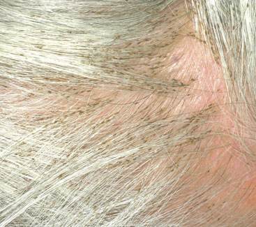
Pediculus humanus capitis (head lice) infection is common in children but can affect people of any age. In this case, the patient presented with a itchy scalp, and on examination was found to have a severe infestation of lice, which had probably been present for several weeks, as the eggs (nits) are attached to the hair some distance from the scalp. Although head lice are not associated with disease transmission, their bites can be extremely irritating (due to allergy to the saliva of the louse) and excoriations can become infected. It is essential to kill both lice and eggs to be sure of successfully eradicating an infection, either by repeating treatment or using a treatment specifically formulated to kill the eggs as well as adult lice.
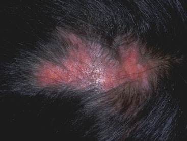
SCALP PSORIASIS
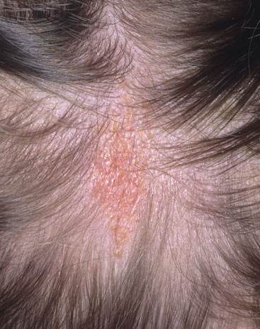
The scalp is one of the common areas to be affected by psoriasis, and indeed may be the only area affected. The skin is usually red with a slivery scale on top. Common places to find it include above the ears and the occipital region. The frontal region may be affected too, but often with less scale - as shown in the picture. It may extend outwards beyond the hairline. The scale may fall and so it is important to wear light coloured clothing to avoid embarrassment. The condition may be confused with seborrhoeic dermatitis (dandruff) but the scale is much thicker in psoriasis. Treatment can be with compounds such as coal tar solution, salicyclic acid, precipitated sulphur in a coconut oil (cocois). Alternatives include a combination of calcipotriol and betamethasone (Xamiol) which is formulated as a gel, arachis oil and coal (Polytar) tar and pure coal tar extract (T/Gel)
NAEVUS SEBACEOUS
This lesion was noticed within a few days of birth. It was an orange-yellow patch, which felt waxy in texture with no hair growth on it. As an adult, the lesion may become more bumpy in nature (verrucous) and is prone to producing benign skin lesions. However, occasionally they may show malignant potential with the development of squamous cell carcinoma or basal cell carcinoma. The parents of the child require counselling about the risks and whether prophylactic excision is appropriate.
ALOPECIA AREATA
Alopecia areata describes patches of hair loss caused by an auto-immune process. It most commonly affects people between the age of 10 and 30 years, and affects both sexes equally. The skin of the scalp will look normal, suggesting the potential for hair regrowth. It possibly occurs more frequently in people who have Down's syndrome and thyroid problems. Closer inspection of the hair may show exclamation mark hairs - short hairs that are narrower at the base. The hair loss may extend to the beard area, eyebrows and eyelashes. Complete hair loss from the scalp may develop (alopecia totalis). There may be nail pitting. After a few months, the hair may re-grow with a white colour initially. Poor prognostic factors include: alopecia totalis, presence for more than 5 years, and development prior to puberty

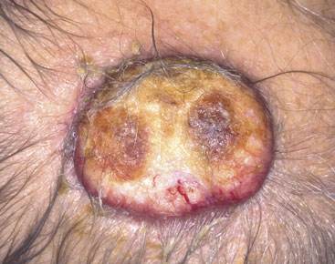
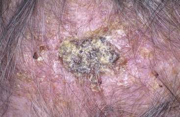
SQUAMOUS CELL CARCINOMA (SCC)
SCCs usually develop where there has been sun damage, so the bald male scalp is a high risk area. Actinic keratoses are a risk factor for the development of SCCs. Bowen's disease, seen more commonly on the legs, is also a fore-runner of the condition. Exposure to radiation and chronic ulcers are also risk factors. Transplant patients are also more at risk a result of immunosuppression and infection with human papilloma virus.
The appearance of this lesion is not typical as SCCs are usually crusty or nodular in appearance. The lesions are usually excised with this one requiring a skin graft. Radiotherapy can be an alternative treatment.
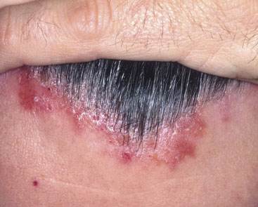
SCARRING ALOPECIA
Alopecia describes hair loss, and one of the prime considerations when making the diagnosis is whether there is any scarring of the skin or not. In this picture, the skin does not appear normal and so it is likely that the hair follicles have been damaged and would be considered scarred. In this case the hair will not re-grow. There may be redness of the skin, which may be encountered in lichen planus and discoid lupus erythematosus. It may be helpful to look on the skin elsewhere to see if there any skin changes that will help with the diagnosis. Pustules may be seen in folliculitis decalvans. Conditions such as shingles may cause scarring as well as a kerion caused by tinea infection. Biopsy may be required to make the diagnosis where there is uncertainty. (See Practice Nurse, 24 February. 2012;42(3):24)
ACTINIC KERATOSIS
Solar keratoses are hard areas of skin that have developed in skin exposed to ultraviolet light long-term. They are more likely to occur in people with fair skin and blue eyes (Fitzpatrick skin type). In primary care, they are most commonly encountered on the scalp of balding men. When they are small, the rough skin that they create may be easier to feel than to see. They may be found at the base of a cutaneous horn and so these lesions should be removed. Alternatively an SCC may be found. The most important consideration is that an actinic keratosis may develop into an SCC. Potential treatments include cryotherapy, curettage and cautery, excision, topical application of 5-fluoruracil or diclofenac 3% gel and photodynamic therapy.
Related articles
View all Articles