Fungal infections
Dr Mike Wyndham
Dr Mike Wyndham
is a GP in a suburban practice in Edgware Middlesex
He has always been a keen photographer, and has been taking medical photographs for more than 25 years. He has been a regular contributor to the GP press and his work has also appeared in the British Medical Journal, The Pharmaceutical Journal and various medical text books
There is an astonishing range of fungal infections, with different appearances and presentations. Management needs to be tailored to the fungus causing the infection
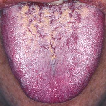
Candidiasis Mouth
Candida albicans is commonly found as a mouth commensal and by definition does not cause any problems. However, people using dentures are more prone to the infection, as are people who have been treated with antibiotics. A suppressed immune system, resulting from chemotherapy or conditions such as HIV disease, is also a risk factor and approximately 90% patients suffering from AIDS will have the infection. People with severe diabetes are more prone, the malnourished and babies may develop the problem too. The infection appears as white spots, which may merge together to form plaques that have a white/yellow colour. Sometimes the condition manifests as a glossitis, a red, sore tongue. Treatment is directed to 'correcting' any underlining condition. The fungus itself can be treated with miconazole gel applied to the affected areas or nystatin suspension. The medication should be swallowed which will treat any associated oesophageal candidiasis. The patient should be recommended not to eat or drink for 30 minutes after treatment. For more severe infections oral fluconazole may be used. It should be remembered that miconazole oral gel is not licensed for use in babies aged under 4 months.
Kerion
Hair loss in an adult is highly unlikely to be caused by fungal infection while in a child there is a strong possibility. It may present as: patches of diffuse scaly skin which may be confused for seborrhoeic dermatitis, well demarcated patches of alopecia with crusting and broken off hairs, a kerion which is a boggy, soft tissue swelling in the scalp. In this image there are pustules present too. The commonest ages affected are from 4-6 years. The organisms that cause the infection are from the trichophyton and microsporum species with the source coming from human, animal or soil. Trichophyton tonsurans has become an increasingly likely cause in recent years particularly affecting African communities in cities. It is helpful to collect scrapings for culture and where this is not possible, samples may be collected with a disposable toothbrush. It is important to be aware that in T.Tonsurans infection, close family members may be carriers. Treatment should be oral, and griseofulvin would be a good choice.

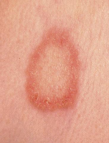
Tinea Corporis
Tinea corporis is also known as ringworm of the body. When communicating with patients, if you use the latter term, ensure that you tell them that the infection is not caused by a worm. Animals such as dogs or cats may act as a reservoir. The author encountered a lesion on the arm of a child that had allowed a hedgehog to walk up and down on the limb! The lesion takes on an annular appearance with a prominent growing edge, which may be scaly and a central area of skin that looks normal (central clearing). Animal ringworms seem to create a more reactive border. Lesions may be single or multiple. Treatment with an imidazole cream will be curative.
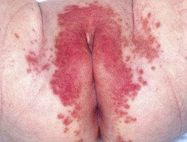
Nappy Rash
An infant's nappy area is constantly exposed to irritating substances and so it is not surprising that from time to time we encounter children with a sore bottom. Fungus likes dark warm moist environments and so the nappy area is ideal. Once an irritant dermatitis has developed, it is an ideal opportunity for candida to proliferate. It appears as red papules extending outward from the dermatitis. Exposing the nappy area to the air is a standard recommended treatment. Parents will then have to take measures to protect the immediate area - furnishings etc - from the baby (particularly for boys!). Personally, I don't recommend this as it may have practical issues. Particularly where the skin is inflamed and may be upsetting the baby, I suggest the application of a combination cream containing hydrocortisone and an anti-fungal agent. I will then suggest applying a standard barrier cream on top. Some practitioners would suggest that this creates a similar environment to the one that caused the problem. My experience is that while that may be true, this management strategy works!
Onychomycosis
When you examine feet for diabetic checks, it becomes apparent how frequently people have dystrophic nails. Many of them will have fungal infection (onychomycosis). Toenails will be infected with dermatophytes such as Trichophyton rubrum. In the elderly, there may be dystrophy without fungus infection as a sign of their age. Psoriasis may cause nail change. In fungal infection, there may be associated skin change in the feet, such as white peeling skin or powdery skin creases. It is worthwhile asking the patient to provide a generous amount of nail clippings to confirm the diagnosis. Treatment with topical application on the nails is not effective and so oral treatment is recommended e.g. terbinafine

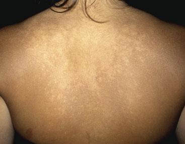
Pityriasis Versicolor
Pityrosporum species, e.g. pityrosporum ovale are normal skin commensals. Their proliferation to becoming pathological is increased by a hot environment and exposure to agents such as suntan lotions. The infection becomes visible when the body is suntanned as the infected areas do not tan and will look lighter than the surrounding skin. The condition may also present itself as brown patches. The front and back of the chest are the commonest areas affected. Treatment depends on the extent of the condition. It should be remembered that the infecting agent is not a dermatophyte (it's a yeast) and so terbinafine will not work. Topical imidazoles such as miconazole or clotrimazole should be applied for 2 weeks. Ketoconazole shampoo can be used. This should be made into a lather, left applied to the skin for 10 minutes and then washed off. This treatment should be carried out four times a week for 2 weeks. For those who wish to take an oral preparation, itraconazole 200mg for 7 days is recommended. The patient should be informed that if they have skin depigmentation from the condition, it will take several months for the skin colour to return to normal.
Related articles
View all Articles