ENT conditions
Dr Mike Wyndhamis a GP in a suburban practice in Edgware Middlesex. He has always been a keen photo...
Dr Mike Wyndhamis a GP in a suburban practice in Edgware Middlesex. He has always been a keen photographer, and has been taking medical photographs for more than 25 years. He has been a regular contributor to the GP press and his work has also appeared in the British Medical Journal, The Pharmaceutical Journal and various medical text books
As this group of images shows, conditions affecting the ear, nose and throat often have visible manifestations — but it is important to make a careful, visual examination to distinguish between conditions with similar presentations
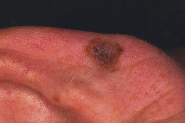
MELANOMA
Observation is probably one of the medical profession's most important skills. My concentration was distracted by the appearance of a pigmented lesion on the patient's ear. The background story was that he had been exposed to a great deal of sun in the early years of his life. When checking for melanoma the ABCD rules should be used. A=asymmetry where the two halves of the mole are different. B=border and here irregular, notched edges are looked for. C=colour. Some normal moles may have two pigments but three is very suspicious. D=dimensions and a check for change in size is important. Inspection of the mole showed that it was a little asymmetrical but not dramatically so. The border was fairly smooth. However, there were at least 3 brown pigments and the patient informed me that it had been growing. There also appeared to be a slight break in the skin in the upper part of the mole, which was not due to trauma. This turned out to be a melanoma in situ and so once removed no follow-up was required.
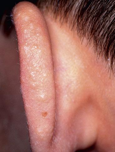
JUVENILE SPRING ERUPTION
This condition is thought to be a variant of polymorphic light eruption where papules and vesicles develop about 24 hours after exposure to the sun. In Juvenile Spring Eruption (JSE), a vesicular eruption appears on the top of the ears of boys between the age of 5-14 years, about 2 hours after exposure to sunlight. It may be recurrent. Symptoms include itch and pain, which may lead it to be confused with herpes simplex. Prevention with the application of sun cream is the best choice of treatment but if it does occur application of 1% hydrocortisone may be helpful.
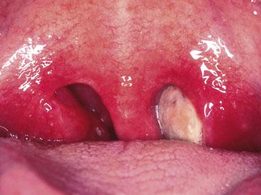
INFECTIOUS MONONUCLEOSIS
Infectious mononucleosis (glandular fever) is caused by the Epstein-Barr virus (EBV) and is characterised by the triad of fever, sore throat and lymphadenopathy. The infection is transmitted by body fluids, with saliva the most well known. However, the virus may be found in secretions from the cervix. The infection is often associated with teenagers, however, I have encountered it in someone as young as 18 months. While the tonsils in glandular fever may have the appearance of a follicular streptococcal infection, they sometimes will look as in this image with a sloughy covering, which is a Dover white colour.
SINUSITIS
Acute sinusitis usually develops after a viral upper respiratory infection. People who are more prone to the infection may have, for example, asthma, cystic fibrosis, nasal polyps or allergic rhinitis. Symptoms include facial pain over the affected sinus, a fever and a purulent nasal discharge. The majority of episodes resolve without requirement for antibiotics. However, if the symptoms are severe or if infected discharge is present for more than 7 days, or are associated with risk factors, then antibiotics are appropriate. Treatment should be with amoxycillin or in patients allergic to penicillin, doxycycline or clarithromycin.
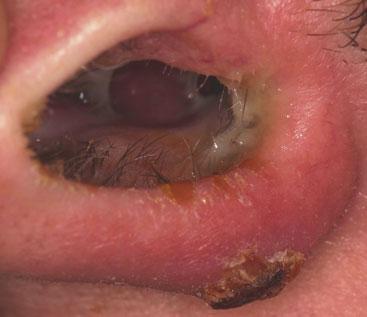
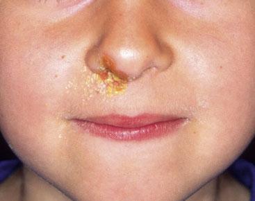
IMPETIGO NOSE
The appearance of bright yellow in any skin rash should suggest infection with Staphylococcus Aureus. The word 'aureus' originating from the Latin word 'aureum' which means gold. Impetigo may also be caused by the Streptococcus. The nose may act as a reservoir for the infection and be perpetuated by the sufferer picking at the crusty rash. Skin conditions such as eczema may be colonized with Staph. Aureus and can predispose to the development of impetigo. If the problem is in the nasal area as shown, I use mupirocin nasal ointment, which should be applied with a cotton bud to prevent spread of the infection.
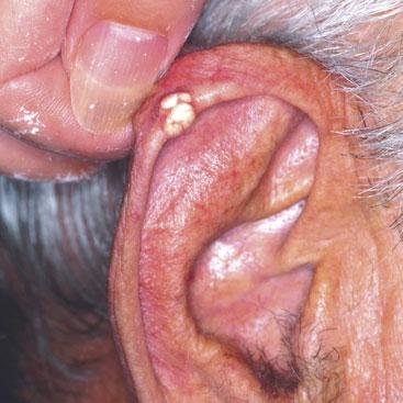
TOPHACEOUS GOUT
Uric acid deposited in the first metatarso-phalangeal joint is our commonest experience of acute gout. However, it may also affect soft tissues such as bursae e.g. the olecranon and also tendons. Uric acid may be deposited in soft tissues and the ear is one of the common places for this to occur. Deposits should not be removed unless there is a complication. Treatment is with allopurinol, which should be co-prescribed in the first month with an oral anti-inflammatory agent or colchicine.
Related articles
View all Articles