Less common rheumatological diseases: an introduction
Sue Brown
Sue Brown
MSc RGN NIP
Consultant Nurse in Rheumatology (Connective Tissue Diseases)
Debbie Bond
MSc RGN NIP
Lead Biologics Specialist Nurse
Nicola Waldron
MSc RGN NIP
Rheumatology Specialist Nurse
All staff at the Royal National Hospital for Rheumatic Diseases NHS Foundation Trust, Upper Borough Walls, Bath
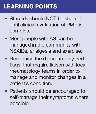
Closer collaboration between practice nurses and rheumatology nurse specialists can heighten awareness of, and improve the diagnosis and treatment of the rarer rheumatological diseases: polymyalgia, ankylosing spondylitis, psoriatic arthritis and systemic lupus erythematosus
Rheumatological diseases are commonly seen in general practice. This article introduces you to polymyalgia rheumatica (PMR), ankylosing spondylitis (AS), psoriatic arthritis (PsA) and systemic lupus erythematosus (SLE). It provides you with information about managing these diseases and discusses the recommendations for the detection and management of the 'red flags' that may need more immediate advice from the local rheumatology team.
POLYMYALGIA RHEUMATICA
PMR is the most common inflammatory rheumatic disorder in people over the age of 50. It is commonly managed in general practice settings. Genetic and environmental factors are thought to contribute to its development although its true causes are, as yet, unknown.1 Typical presentations include neck, shoulder, low back and thigh pain that improve on exercise. It is associated with early morning stiffness (lasting 45 minutes or more) and raised inflammatory markers.
Symptoms are similar to rheumatoid arthritis (RA) and spondyloarthropathies and the onset of symptoms can be acute or gradual.2 For some, the diagnosis is straightforward but, for those with atypical presentations, diagnosis can be difficult.3 PMR also shares many of the pathogenic features of giant cell arteritis (GCA); a common large vessel vasculitis also seen in those over 50 years and which can sometimes appear later.4 Up to 5-10% of people with PMR are also diagnosed with GCA. It is therefore essential that PMR is correctly diagnosed. GCA, infection and cancer, as a cause for the symptoms and serological changes, should be screened for before PMR is confirmed.
Treatment and management
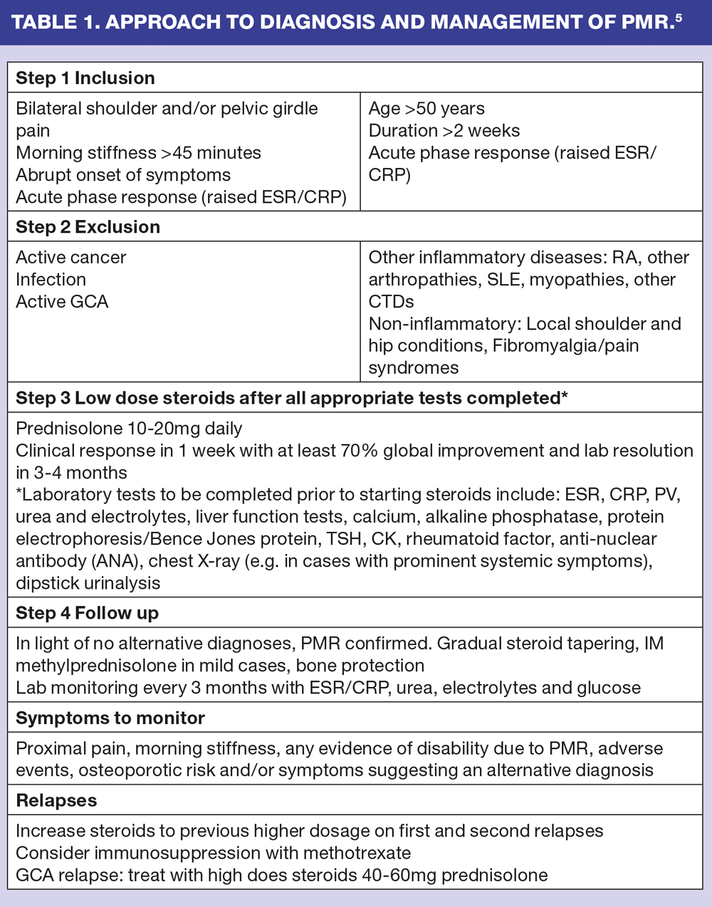
Guidelines for the management of PMR have been developed to improve early diagnosis and initiation of appropriate treatment.5 These include advice that:
- Unless GCA is the clear diagnosis, urgent initiation of steroids is not required until clinical evaluation is complete
- A stepped approach to diagnosis, treatment and management is essential. (Table 1)
Corticosteroid treatment usually lasts for 1-2 years. The guidelines recommend a starting dose of 15mg oral prednisolone daily for 3 weeks (with bone protection). The dose should then be reduced by 2.5mg for 3 weeks. 10mg is then given for 4-6 weeks, with slow tapering thereafter of 1mg every 4-8 weeks or alternate day reductions. Clinical response is usually seen within the first week of treatment. Treatments must be tailored to the individual and some may benefit from a more gradual reduction regime. Intramuscular methylprednisolone can reduce the longer term risks associated with oral corticosteroids.
Referral to secondary care for specialist advice is recommended by the British Society of Rheumatology (BSR) for people with atypical features of PMR,5 especially in the following situations:
- Younger age (usually under 60 years)
- No shoulder involvement
- Lack of inflammatory stiffness
- Insidious onset
- Normal or very high inflammatory markers
- Red flag features (prominent systemic features, weight loss, night pain, neurological signs)
- Suspicion of co-existent GCA
- Poor or incomplete response to corticosteroids
- Difficulty reducing corticosteroid dose
- Recurrent relapse
- Contraindication to corticosteroid treatment.
ANKYLOSING SPONDYLITIS
AS is an inflammatory spondyloarthropathy affecting the spine and sacroiliac joints (SIJ). It affects up to 1% of the population depending on race, with less than 1% of non-Caucasians affected, and is more frequently seen in males.6
Individuals with AS experience back pain and stiffness. Inflammation and damage to the joints can cause narrowing or fusion of the SIJ and can cause ankylosing (fusion) of the vertebra giving the classic, X-ray appearance of 'bamboo spine'.(Figure 1) Peripheral joints, such as knees, hands and feet may also be affected, leading to people being misdiagnosed with other inflammatory conditions such as RA. AS carries a high risk of other associated conditions, such as acute anterior uveitis. This can affect up to 40% of people with AS and inflammatory bowel diseases.6 In common with other inflammatory conditions there is also a higher risk of cardiovascular disease.7
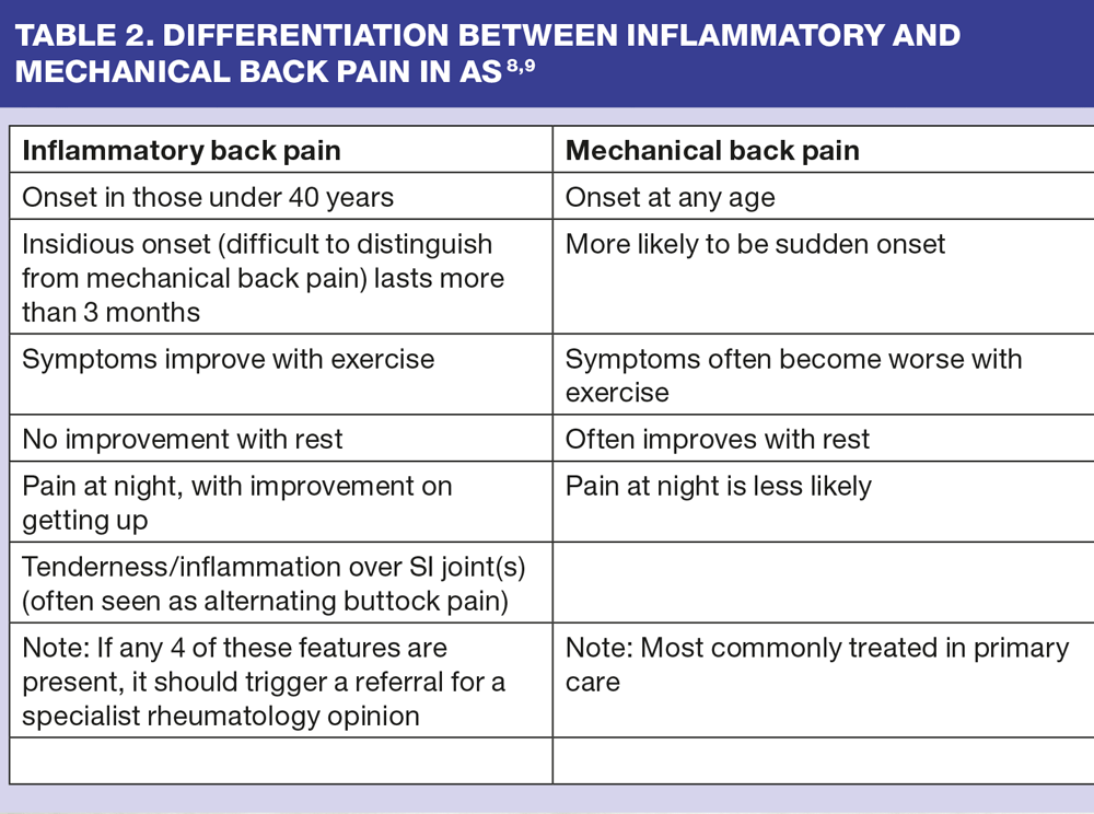
Early identification of inflammatory back pain is important in preventing long term joint destruction. However, one of the major factors causing diagnostic delay is difficulty in differentiating between inflammatory and mechanical back pain. The information in table 2 can help you decide whether the patient needs a specialist referral to rheumatology.8,9
Once inflammatory back pain is identified, diagnosis can, however be further delayed as current diagnostic criteria use X-ray changes which can take 5 years or more to become evident. The use of magnetic resonance imaging (MRI) can show changes in the SIJ at an earlier stage than can be detected by plain X-ray.10
Signs and Symptoms of AS include:
- Early morning stiffness, which can last 2 hours or more
- Pain more associated with rest than activity
- Poor sleep — waking in the early hours with back pain
- Enthesitis (inflammation at the site of insertion of a tendon or ligament) is common around the heel and Achilles tendon 6
- Restriction in movement in the spine and/or chest
- A pattern of flaring and remission
Treatment and management
Non steroid anti-inflammatory drugs (NSAIDs) are the main drugs used to control symptoms and they may have a disease modifying action in AS. A proton pump inhibitor (PPI) should be co-prescribed if NSAIDs are to be used long-term. Unless there are peripheral symptoms the use of disease modifying anti-rheumatic drugs (DMARDs) is of limited benefit.11 Most patients are managed with NSAIDs and PPIs and may not be seen at the surgery very often.
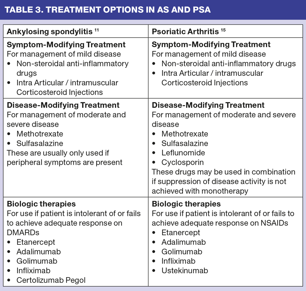
The most effective, currently used, biological treatments for AS are anti-tumour necrosis factor (anti-TNF) agents. These target tumour necrosis factor, the pro-inflammatory cytokines responsible for the establishment and maintenance of inflammation in many rheumatological conditions. These biologic treatments can only be prescribed if the individual meets NICE criteria.12 Biologics are also used in PsA: this is discussed in the next section and Table 3 summarises treatment options in AS,12 and PsA.13,14
Exercise is an important management tool in AS.11 Patients can attend National Ankylosing Spondylitis Society (NASS)-led exercise sessions, which can include hydrotherapy, and which are usually supported by physiotherapists. The combination of pharmacological treatments and regular exercise are most effective and can enable the individual to live a normal life.
PSORIATIC ARTHRITIS
PsA is an inflammatory arthritis associated with psoriasis.16 Although any joint can be involved, commonly the peripheral joints, such as feet, hands, ankles and shoulders, are affected. Patients may or may not present with skin psoriasis, but for a positive diagnosis there must be a history of psoriasis, first-degree family history or evidence of nail psoriasis. Extra-articular features can include iritis, uveitis and inflammatory bowel disease.
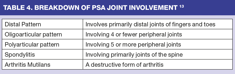
Patients may present with a range of disease severity, from mild disease — where just one joint is affected — to the more destructive form with accompanying poor prognosis. Table 4 gives a breakdown of joint involvement in PsA.15
Treatment and management
Left untreated, a substantial proportion of patients develop persistent inflammation leading to progressive joint damage and functional disability.17 Treatment goals are similar to all rheumatic diseases, i.e. reducing pain, stiffness and swelling, inhibiting disease progression, and helping the patient maintain a reasonable quality of life. The drug groups currently used in the management of PsA are similar to those used in AS, and include DMARDs, biologic therapies, NSAIDs and intra-articular or intramuscular corticosteroids.
Patients with mild disease may be managed with NSAIDs alone, and injections of corticosteroids when required, although it is important to note that corticosteroid use may flare their psoriasis. However, these treatments will not inhibit the development of structural joint damage.18 If the disease is more active DMARDs should be introduced. All but sulfasalazine may have a positive impact on a patient's psoriasis as well as moderating joint activity. For those who are either intolerant of or fail to respond to selected DMARDs, one of the biologic therapies will be considered.16
SYSTEMIC LUPUS ERYTHEMATOSUS
SLE (or lupus) is a rare, multi-system, autoimmune disease of unknown aetiology. It commonly affects the skin, joints and mucous membranes and, less commonly, can become a multi-organ disease that results in renal, haematological and neurological complications.
SLE is difficult to diagnose due to the insidious nature of symptoms and its rarity in general practice settings.19 Autoantibodies can be measured to aid diagnosis; these include ANA (anti-nuclear antibody), dsDNA (double stranded deoxyribonucleic acid), ENA (extractable nuclear antigen), and especially the anti-nuclear autoantibodies anti Sm, anti Ro/SSA and anti La/SSA. In pregnancy the anti Ro/SSA antibody can be associated with a small risk of neonatal lupus in the child, including congenital heart block (5% risk), neonatal transient skin rash (2%) and haematological and hepatic abnormalities.20 Outcomes of SLE in pregnancy, however, are now significantly improved by multi-disciplinary management.
SLE is a worldwide disease and is seen more commonly in women than men, with approximately 10 women diagnosed during child bearing years for every one man diagnosed.21 The diagnostic classification criteria recommend that 4 out of 11 criteria need to be met. (Table 5)22 Lower socio-economic status and Asian and Hispanic ethnicity is linked to a greater risk of complications, such as renal disease, and higher mortality rates.23
Treatment and management
SLE can vary in its intensity, with significant variance in symptoms between individuals. With the exception of an extremely mild form of SLE that is in remission, all patients should be managed under shared care arrangements between the hospital and general practice.
Early diagnosis is vital in helping to prevent serious complications. The most important message for primary care is to maintain a high level of vigilance, especially in a young female patient experiencing any of the more commonly seen symptoms. These include:
- Severe fatigue
- Flitting arthralgias and myalgias
- Mouth ulcers
- Headaches
- Rashes, particularly provoked by the sun
- Persistent proteinuria
- Worsening symptoms during and prior to menstruation.
All patients should be assessed for the presence of antiphospholipid antibodies which may predispose them to thromboembolic events such as pregnancy loss, deep vein thrombosis or pulmonary embolus. Once detected, this can be treated with low dose aspirin (in the absence of any thromboembolic events) or long term warfarin if the patient has already experienced a DVT or PE.24
Hydroxychloroquine is an important drug therapy for SLE, unless side effects prevent its use. It has mild disease modifying properties, but, importantly, there is evidence of cardio-protective qualities and improved outcomes in pregnancy.25,26 The use of corticosteroids, as either intramuscular or intravenous injections, or higher doses orally, can be life-saving. Oral corticosteroid doses will be influenced by disease activity and risk of or worsening of systemic involvement, and should always be reduced as soon as possible. Further immunosuppression with azathioprine may be required for general SLE symptoms, methotrexate for more joint related symptoms and mycophenolate mofetil if disease activity remains uncontrolled. Induction with intravenous cyclophosphamide is always used if there is evidence of systemic involvement. Treatment plans must always be negotiated and agreed with the patient in order to ensure concordance with essential medication.
THE ROLE OF THE PRACTICE NURSE
There are many ways in which you, in collaboration with specialist, secondary care teams, can be involved in the care of patients with PMR, AS, PsA and SLE, and many ways in which you can positively influence how these individuals manage and live with these long-term conditions.
DMARDS and biologics
Patients on DMARDs and biologics require regular blood monitoring (see BSR monitoring guidelines) and this is frequently carried out in general practice. Any significant abnormalities should be reported to the patient and their rheumatology team so that appropriate action can be taken.
Flares
You are well placed, should a patient present with an exacerbation (flare) of their symptoms, to give advice about:
- Rest
- Taking prescribed analgesia and anti-inflammatory medication
- Taking short-term corticosteroid therapy, if appropriate.
Longer-term pain management
Patients may require support and advice about the pharmacological and non- pharmacological options for longer-term pain management. This will include the appropriate use of NSAIDs alongside regular analgesics and alternative interventions such as TENs machines and hot wax therapies. Heat and cold packs can offer much relief.27 Referral to physiotherapy will sometimes help to maintain joint ranges and improve function by strengthening muscle groups. You can also direct patients to further sources of information, such as Arthritis Research UK. (www.arthritis-researchuk.org.uk)
Fatigue
Patients with rheumatological diseases often experience high levels of fatigue — a constant feeling of exhaustion not improved by rest or sleep — and for some, this is more debilitating than the joint symptoms.28 You can work in collaboration with other specialist therapists to enhance the patient's ability to manage fatigue, and reduce their frustration. Of particular help is advice about:
- Sleep
- Pacing
- Goal setting
- Relaxation techniques.
Fatigue can be a particularly significant problem in lupus. Referral back to secondary care, for intensive occupational and physiotherapy, may be necessary.
Corticosteroid side effects
Monitoring for side effects of corticosteroids is an important aspect of your role. All patients, especially women, should have an osteoporosis risk assessment and, when indicated, start calcium and vitamin D replacement and bisphosphonate therapy. A referral for a DEXA scan is imperative in those with previous or high fracture risk, and those with a family history of osteoporosis.
Vaccination advice
Patients who are immunosuppressed with high dose corticosteroids, DMARDs and biologic treatments should avoid all live vaccines. Flu and pneumococcal vaccines are safe and recommended as these patients are at higher risk of infections. If you are in doubt, liaise with the rheumatology team.29
Urinalysis
Simple urinalysis, when bloods are due, can alert you to the presence of early renal disease in SLE. This is particularly important when blood pressure, urea, creatinine and estimated glomerular filtration rate (eGFR) show decline in kidney function. A urinary protein: creatinine ratio at this stage is vital to further quantify any significant protein leak signifying renal complications which may need urgent treatment and early referral back to secondary care.
Cardiovascular risk
Accelerated atherosclerosis is a major cause of morbidity and mortality in inflammatory rheumatological disease. 30 (Table 6) Giving advice and supporting people to reduce their cardiovascular risk is already a major part of the primary care workload and many practice nurses have considerable expertise in this area. Advice, continuing support and monitoring of risk is essential for those with SLE and other inflammatory diseases.
Pregnancy
For patients with SLE, pregnancy requires full collaboration between the individual and their primary and secondary care teams in order to optimise outcomes for both mother and baby. This is especially important for those women who have had previous multi-system involvement and carry antibodies that may confer greater risk. Serious SLE complications can develop rapidly. Quick and easy access to specialist services able to manage and treat flares is vital.
Red flags and access to hospital services
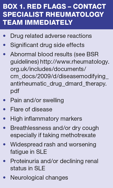
Although patients will be reviewed regularly at their local rheumatology unit, you will often be the first port of call for someone experiencing an exacerbation of symptoms. Close collaboration with your local rheumatology specialist nurse team should be encouraged and will enhance the support you are able to offer these patients. This is especially important if you identify a 'red flag'. (Box 1)
SUMMARY
Managing patients with any rheumatological disease can be challenging, but hugely rewarding. These patients have often endured delays in diagnosis, sometimes stigmatising skin diseases, as well as many years of difficult symptoms and disabling joint pains. This places a psychological and physical burden on the individual that requires effective collaboration between primary and secondary care to ensure continuity of ongoing and effective support.
REFERENCES
1. Kermani T, Warrington K. Polymyalgia Rheumatica. Lancet 2013; 381: 63-72
2. Nesher G. Polymyalgia rheumatica — diagnosis and classification. Journal of Autoimmunity 2014; 48-49; 76-78
3. Mallen C, Heliwell T, O'Brien A, Mackie S. Polymyalgia rheumatica. Reports on the Rheumatic Diseases. Arthritis Research UK. Spring 2014; Series 7: Hand On No. 4
4. Smeeth L, Cook C, Hall AJ. Incidence of diagnosed polymyalgia rheumatica and temporal arteritis in the UK 1990-2001. Annals of the Rheumatic Diseases. 2006; 6 (8): 1093-8
5. Dasgupta, B., et al. BSR and BHPR Standards Guidelines and Audit Working Group. BSR and BHPR guidelines for the management of polymyalgia rheumatica. Rheumatology (Oxford) 2010; 49: 186-190
6. Gossec L, Dougados M. Clinical Features. In Dougados M, van der Heijde D (Eds.) Ankylosing Spondylitis. 2004. Health Press, Oxford. 32 - 45
7. Bakland G, Gran JT, Nossent JC. Increased mortality in ankylosing spondylitis is related to disease activity. Annals of the Rheumatic Diseases. 2011; 70 (11): 1921-1925
8. Sieper J, van der Heijde D, Landewé R et al. New criteria for inflammatory back pain in patients with chronic back pain: a real patient exercise by experts from the Assessment of Spondyloarthritis International Society (ASAS). Annals of the Rheumatic Diseases. 2009; 68 (6): 784-788
9. Harris C, Gurden S, Martindale J, Jeffries C. Differentiating Inflammatory And Mechanical Back Pain: Challenge Your Decision Making. 2012. www.astretch.co.uk/M208 IBP Module Booklet.pdf
10. Rudwaleit M, van der Heijde D, Landewé R et al. The development of Assessment of Spondyloarthritis International Society classification criteria for axial spondyloarthritis (part II): validation and final selection. Annals of the Rheumatic Diseases. 2009; 68 (6): 777-783.
11. Zochling J, van der Heijde D, Burgos-Vargas R, et al. ASAS/EULAR recommendations for the management of ankylosing spondylitis. Annals of the Rheumatic Diseases. 2006; 65: 442-452
12. National Institute for Health and Care Excellence. Adalimumab, Etanercept and Infliximab for Ankylosing Spondylitis. 2008. Technical appraisal No. 143. NICE, London.
13. National Institute for Health and Care Excellence. Adalimumab, Etanercept and Infliximab for the treatment of psoriatic arthritis. 2010. Technical appraisal No. 199. NICE, London
14. National Institute for Health and Care Excellence. Golimumab for psoriatic arthritis. 2011. Technical appraisal No. 220. NICE, London
15. Gladman D, Rosen C, Chandra V. Psoriatic Arthritis. 2014. Publ: Oxford University Press, pp. 69-74.
16. Gladman D, Chandran V. Psoriatic Arthritis — The Facts. 2009. Publ: Oxford University Press, Oxford
17. Chang A, Gotlieb, AB, Lizzul PF. Management of psoriatic arthritis from the view of the dermatologist. Nature Reviews/Rheumatology. 2011; 7 (10): 558-598
18. Gottleib A, Korman NJ, Gordon KB et al. Guidelines of care for the management of psoriasis and psoriatic arthritis: Section 2. Psoriatic arthritis: overview and guidelines of care for treatment with an emphasis on biologics. Journal of the American Academy of Dermatology 2008; 58: 851-864
19. Hughes G. Lupus — a GP guide to diagnosis: compiled by Yvonne Norton 2000
20. Bramham K, Soh MC, Nelson-Piercy C. Pregnancy and renal outcomes in lupus nephritis: an update and guide to management. Lupus. 2012; 21: 1271
21. Cervera R, Khamashta MA, Font J, et al. Morbidity and mortality in systemic lupus erythematosus during a 10-year period: a comparison of early and late manifestations in a cohort of 1,000 patients. Medicine (Baltimore) 2003; 82: 299—308.
22. Hochberg MC, for the Diagnostic and Therapeutic Criteria Committee of the American College of Rheumatology. Updating the American College of Rheumatology revised criteria for the classification of systemic lupus erythematosus [letter]. Arthritis Rheum. 1997; 40:1725
23. Lau CS, Yin G, Mok MY. Ethnic and geographical differences in systemic lupus erythematosus: an overview. Lupus. 2006; 15:715—9.
24. Ruiz-Irastorza G, Cuadrado MJ, Ruiz-Arruza I et al. Evidence-based recommendations for the prevention and long-term management of thrombosis in antiphospholipid antibody-positive patients: Report of a Task Force at the 13th International Congress on Antiphospholipid Antibodies: Lupus. 2011; 20: 206
25. Tang C, Godfrey T, Stawell R, Nikpour M. Hydroxychloroquine in lupus: emerging evidence supporting multiple beneficial effects. Internal Medicine Journal. 2012; 42: 9, 968-978
26. Izmirly PM, Kim MY, Llanos C, et al. Evaluation of the risk of anti-SSA/Ro-SSB/La antibody-associated cardiac manifestations of neonatal lupus in fetuses of mothers with systemic lupus erythematosus exposed to hydroxychloroquine. Annals of Rheumatic Diseases 2010; 69: 1827—1830.
27. Oosterveld FGJ, Rasker JJ. Treating Arthritis with locally applied heat or cold. Seminars in Arthritis and Rheumatism. 1994; 24 (2): 82 — 90
28. White, C. Fatigue and Sleep in Rheumatology Nursing — A Creative Approach. Ed Jackie Hill Publ: John Wiley & Sons Ltd. 2006. Chichester 2nd Edition pp 24
29. British Society of Rheumatology. Vaccinations in the immunocompromised person. Guidelines for the patient taking immunosuppressants, steroids and the new biologics therapies. 2002. http://www.rheumatology.org.uk/includes/documents/cm_docs/2009/v/vaccinations_in_the_immunocompromised_person.pdf
30. Wajed J, Ahmad Y, Durrindton PN, Bruce IN. Prevention of cardiovascular disease in systemic lupus erythematosus"”proposed guidelines for risk factor. Rheumatology (Oxford) 2004; 43: 7-12.


Related articles
View all Articles