Tuberculosis: a clear and present danger
Dr Mary Lowth
Dr Mary Lowth
MA MB BChir FRCGP PGMedEd
Tuberculosis, along with HIV, is a leading cause of death worldwide. While most deaths occur in low- and middle-income countries general practice nurses need to be aware and alert to its presence in the UK
Tuberculosis (TB) is the second leading infectious disease killer worldwide.1 Despite major advances in tackling its spread, about one third of the world’s population has latent TB.2,3
Most TB deaths occur in low and middle income countries, but it has never left our shores, and practice nurses need to be aware of, and alert to its presence. Particular risk groups in the UK include refugees and asylum seekers, recent immigrants from endemic regions, homeless people, problem drug users, and prisoners.
GLOBAL CONTEXT1–3
The World Health Organization’s (WHO) Millennium Goals, set in 1990, aimed to reverse a relentless increase in TB deaths. TB mortality has fallen by nearly half since then, but we cannot be complacent. WHO believes that 1 in 3 of all living humans (2 billion people) are infected with TB. In most of these people the TB is latent.
In 2014 9.6 million people were estimated to have acquired TB, and 1.5 million people died of it (of whom 0.4 million were HIV positive). Of these deaths, 140,000 were in children, and nearly half a million were multi-drug resistant.
About 80% of people in many Asian and African countries test positive to the tuberculin test, while 5–10% of people in the United States population tests positive.
From 2016 the WHO’s new goal is to cut TB deaths by 90% and new cases by 80% by 2030.
UK EPIDEMIOLOGY
Public Health England last reported on TB in the UK in 2014, when there were 6,520 new diagnoses.4
Of these, 73% occurred in people born outside the UK (15% were among recent migrants). The rate of TB among the non UK-born population was almost 20 times the rate in the UK-born, at 80 per 100,000, although it is declining. In the UK-born population rates remain stable at 4.1/100,000 per year. Those most at risk remain:
- Individuals from ethnic minority groups
- Those with social risk factors
- The elderly.
For 28%, treatment started more than four months after onset of symptoms.
There were 12.3 new cases per 100,000 population, meaning that, on average, a practice with a list size of 10,000 would see a new case every year. However, the spread is not even.
Most TB cases occurred in large urban centres, among young adults, those from countries with high TB burdens, and those with social risk factors for TB. Some 70% of cases lived in the 40% most deprived areas.
London had the highest proportion of cases in the UK (37.8% of all cases, with an incidence of 35.5 cases per 100,000), followed by the West Midlands (17.3 per 100,000).
More than 7% showed resistance to any first-line drug and 1.6% were multidrug-resistant.
TB AND HIV COINFECTION1-3
TB is the most common opportunistic infection in people living with HIV worldwide. It is also the most common cause of death in HIV positive adults. Where both HIV infection and latent or active TB are present, each disease speeds up the progress of the other.
In 2014, of the estimated 9.6 million people who developed TB, 13% were HIV positive. Around 25% of all TB deaths, and around 25% of the estimated 1.2 million deaths from HIV/AIDS were patients with coinfection.
THE TB BACTERIUM
Mycobacterium tuberculosis is a rod-shaped, aerobic bacterium. It has an unusually waxy cell wall, which makes it acid fast (meaning it can hold dye when exposed to alcohol) – hence the name ‘acid-fast bacillus’ or ‘AFB’ on a lab test.
WHERE TB CAME FROM5,6
TB evolved early, side by side with mankind and has been shadowing us for at least 70,000 years. The diversity of the mycobacterium increased when human populations expanded, and an increasingly deadly form evolved. Highly infectious and lethal, it was also able to survive in low population densities, using periods of latency followed by reactivation.
LATENT VS ACTIVE DISEASE
Of the 2 billion people in the world who have TB most have latent disease, meaning that they are asymptomatic and non-infectious. The bacterium is ‘locked away’ inside inflammatory lesions in the body – usually the lungs (see below).
Moving from latent to active disease occurs because of host debilitation. Typical causes are:
- Ageing
- Coexisting disease (particularly HIV but also diabetes, chronic kidney disease, inflammatory diseases, cancers)
- Nutritional deprivation
- Immunosuppression, e.g. with oral steroids or chemotherapy.
Reactivation usually occurs at the lung apices. A person with active infection has replicating TB organisms. The level of infectiousness varies with the stage and site of their infection, but patients with active pulmonary TB may cough up huge numbers of mycobacteria and spread the infection.
PATHOGENESIS AND PATHOPHYSIOLOGY
Acquisition of infection
The organism is usually acquired through inhalation of infected material. Most organisms we inhale are captured by the mucus of the large airways, and those that make it down to the deep alveoli are absorbed into the blood stream and destroyed by white blood cells (phagocytes and macrophages). However, in the case of mycobacterium tuberculosis the phagocyte swallows the bacterium, but does not succeed in killing it. Instead the bacterium proliferates inside the cell. The individual now has primary (latent) TB. At this point most people are asymptomatic, although a few experience a mild flu like illness.
Granuloma formation
After about 3 weeks, slow, ‘cell mediated’ immunity kicks in. This involves T cells, which attack the infected cells. The resulting inflammatory lesion forms a granuloma. The tissue in the middle dies, a process called caseous necrosis, and what remains is called a Ghon focus. This site of primary TB infection is most commonly sub-pleural in the middle to lower zones. At this point one of several things happen:
- The immune system wins and the TB is eradicated
- The walling off of the mycobacterium is complete and spread does not occur, but the mycobacterium remains latent and capable of reactivation
- The infection spreads to the upper lobes, where oxygen is richest.
Rapidly spreading lesions tend to cavitate, which may lead to fast local spread in the lungs and to dissemination via the blood (systemic miliary TB).Calcification in the lesions is common, which usually makes them visible on X-ray.
Miliary TB occurs when the mycobacterium spreads throughout the body in the blood stream. All organs can be affected but those in which TB is most commonly seen are:
- Kidneys – where it causes genitourinary symptoms and sterile pyuria (AFB may be revealed on special staining, but this is not part of a microscopy and culture unless you ask for it)
- Meninges – where it causes TB meningitis, a highly destructive and lethal form with insidious onset
- Lumbar vertebrae – where it causes back pain
- Adrenal glands – where it destroys adrenal tissue, leading to Addison’s disease
- Liver – where it causes TB hepatitis
- Cervical lymph nodes – where it causes lymphadenitis, known as scrofula in medieval times.
RISK FACTORS, 1,7
The main risk factors for TB are as follows:
- Close contact with a TB patient: household members of an infectious case have a 1 in 3 chance of contracting the disease
- Non-UK ethnicity: this group account for 72% of UK TB patients. Most are diagnosed within five years of entering the UK but their lifetime risk remains high
- Homelessness
- Addiction to alcohol or other recreational drugs
- Reduced resistance: impaired immunity, debility, starvation, HIV/AIDs, end stage renal disease, steroid therapy, diabetes, systemic inflammatory disease, cancer
- Increasing age.
Young children are particularly susceptible due to their immature immune systems. They rarely develop cavitating disease and are therefore rarely infectious. Children and the immunosuppressed are more likely to get extra-pulmonary TB.
SYMPTOMS1
These vary depending on the site of infection. If the TB is latent there will be none.
Onset is often insidious. A high index of suspicion is needed for diagnosis.
General symptoms (not always present) include fatigue, malaise, weight loss, anorexia, failure to thrive, night sweats, fever. These may advance very insidiously. Findings may include leucocytosis and anaemia. Most patients in the UK present with cough, general symptoms or a history of contact.
About 60% of UK cases are of pulmonary TB. Classical symptoms are:
- Cough for >3 weeks
- Weight loss/anorexia
- Fever
- Night sweats
- Haemoptysis
- Chest pain
- Fatigue.
The most common site outside the lungs is the kidney. It presents with pyuria (white cells in the urine) without visible organisms, inaccurately termed sterile pyuria. Genitourinary TB is thought to be the cause of 10% of female infertility worldwide.
Musculoskeletal infection causes pain and, occasionally, an abscess detectable as a mass. The lumbar spine is the most common site (Potts disease). Lymph node infection presents with palpable nodes, which may initially be firm and tender but later may suppurate. Pus can be sent to the lab in a dry pot. Erythema nodosum may occur early in the disease process. Other, less common sites include TB meningitis, TB pericarditis and gastrointestinal TB (which can mimic inflammatory bowel disease).
Elderly and immune-impaired individuals may have only very mild symptoms, since many of the symptoms of TB come not from the organism itself but from the host immune response.
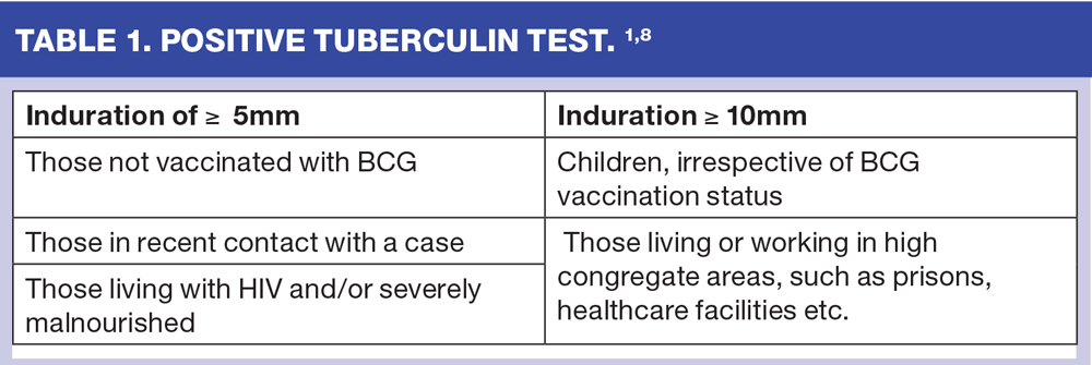
EXAMINATION, INVESTIGATION AND DIAGNOSIS1,8
There may be nothing to find on examination, although upper lobe signs are the classic presentation. Other signs will vary with the site of infection.
Skin testing (Mantoux test)
Tuberculin is injected in between the layers of the dermis, and the reaction is reviewed at 48-72 hours. (Tuberculins are proteins extracted from the bacterium, the most important type being purified protein derivative, PPD). Criteria for a positive test are shown in Table 1.
Skin testing detects exposure but cannot say if infection is cleared, latent or active. It may be positive in individuals who have had BCG, and in the presence of exposure to other mycobacteria.
IGRA (interferon gamma release assay)
This is a more specific blood test for TB, and is not likely to be positive after BCG vaccination. It only detects the presence of mycobacterium tuberculosis and not the Bacillus Calmette-Guérin used in BCG vaccination, or most other non-tuberculous mycobacteria.
Chest X-ray
TB is usually identifiable as an apical pulmonary lesion (in primary TB there may also be a pleural effusion). Miliary TB causes a speckled appearance. A normal Chest X-ray makes pulmonary TB unlikely.
Respiratory TB
Microbiology is needed to detect the organism and to determine sensitivities. Coughed sputum samples (at least three, including one early morning sample) may detect the disease. Bronchial lavage may be used to obtain samples.
Non-respiratory TB
Where sterile pyuria occurs, three early morning urine samples should be sent with a request for examination for AFB. Biopsy and needle aspiration may be needed for solid lesions.
Sending samples, whether sputum, urine or from lesions elsewhere and testing for AFB is the gold standard for diagnosing TB.
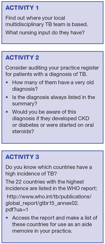
CONTACT TRACING
The local multi-disciplinary TB team will advise on contact tracing. They normally offer Mantoux testing to household contacts and to close work or school contacts (aged 5 years and older) of all patients diagnosed with active TB. IGRA is recommended as a second-line test, and for BCG-vaccinated people. The approach varies slightly in children under 5 years.
MANAGEMENT 1,8-10
If clinical signs and symptoms suggest a diagnosis of tuberculosis, starting treatment without waiting for culture results is usually advised. The standard regimen is then completed even if culture results are negative.
Most cases can be managed as outpatients but occasional admission may be needed (to a single side-room vented to the outside until proven to be non-infectious). TB specialist physicians with access to TB nurse specialists and health visitors should manage all patients with TB, regardless of the site.
Management depends on the sensitivities of the organism. Active TB is treated with combination therapy. The standard regime for all TB is 6 months of isoniazid and rifampicin supplemented in the first 2 months with pyrazinamide and ethambutol. The exception is central nervous system TB, which is treated for 12 months.
Patients become non-infectious in weeks but prolonged treatment is needed to eradicate organisms. Multiple drugs are used to try to prevent resistance, and patients must complete the course.
There are two treatment options recommended for latent TB: isoniazid for six months or a combination of rifampicin and isoniazid (called rifinah) for three months. This treatment is also considered for close contacts of proven cases and those tested positive on contact tracing.
MULTI-DRUG RESISTANT TB 10
Poor chemotherapy over time has led to drug-resistant disease. Drug resistance develops by the selective growth of resistant mutants. The proportion of drug-resistant mutant bacteria in an individual to even one drug is fairly low, and combination chemotherapy with three or more drugs usually results in cure. However, irregular treatment, inadequate drugs, inadequate drug doses or addition of a single drug to a failing regimen allows selective growth of resistant mutants and acquired drug-resistant TB. Contacts of these resistant cases develop primary drug resistant TB.
Drug resistance in tuberculosis is a man-made problem. Anti-TB therapy (ATT) must be given optimally by:
1. Ensuring adequate absorption of drugs
2. Timely diagnosis and management of drug toxicities, and
3. Treatment adherence.
New classes of anti-TB drugs are needed, but are unlikely to become available soon.
NOTIFICATION
All cases of TB must be notified to the local communicable diseases consultant. The doctor suspecting the diagnosis is legally responsible for notification.
PREVENTION OF TRANSMISSION
Adults with reactivated TB are the most infectious, and droplet spread means they can disseminate organisms very quickly. However not all patients with active TB need to be isolated (and not all need to be hospitalised). Management of active cases will need the advice of your local multidisciplinary TB team.8
VACCINATION
The Bacillus Calmette-Guérin Vaccine (BCG) was first used in 1921. It is a live vaccine derived from bovine tuberculosis. In countries with a high prevalence of TB it is given as soon as possible after birth but is absolutely contraindicated in babies with immune deficiency, including HIV.
BCG vaccine prevents only about 20% of infections, but when infection does occur it prevents around 50% from moving from latent to active. It is most effective in preventing miliary TB in children, has little effect beyond the age of 16 years and none beyond the age of 35 years. It is believed to be effective for around 15 years.
Patients over the age of 6 years (unless they have had known contact with TB or have lived/travelled to a high incidence country) are first skin tested to look for evidence of previous exposure. With a positive Mantoux test the local reaction to a BCG immunisation can be severe. BCG is given intradermally.
The UK introduced universal BCG vaccination in 1953, and stopped it in 2005 because of decreased prevalence (the number of vaccines needed to prevent one infection increased from 94 to over 12000.)
It is still given to some health professionals, and to high-risk babies and children, namely:
- Those born in a higher risk area (including some London boroughs)
- Those with a parent or grandparent born in a country with a high rate of TB (See activity 3)
- Children recently arrived from areas with a high incidence
- Children who are contacts of active cases.
PREVENTION1,8,9
NICE Pathways emphasise education and information, both to the public and to healthcare professionals, as the best strategies for awareness-raising and early detection.
SUMMARY
Tuberculosis remains a global threat. In the UK we need to remain alert. This is particularly true if you work in a higher risk area or treat higher risk groups, but none of us can be complacent.
New drugs and vaccines are urgently needed to fight the disease. Multidrug-resistance against first-line treatments is a growing threat in many countries.1,5,10
REFERENCES
1. International Council of Nurses. TB Guidelines for nurses in the care and control of tuberculosis and multi-drug resistant tuberculosis. 2016. 3rd Edition. http://www.icn.ch/images/stories/documents/projects/tb/tb_mdrtb_guideline.pdf
2. WHO Global tuberculosis report 2015 http://www.who.int/tb/publications/global_report/en/ FulL report at http://apps.who.int/iris/bitstream/10665/191102/1/9789241565059_eng.pdf?ua=1
3. WHO Factsheet 104. March 2016 http://www.who.int/mediacentre/factsheets/fs104/en/
4. Public Health England Tuberculosis in the UK 2014 Report: https://www.gov.uk/government/uploads/system/uploads/attachment_data/file/360335/TB_Annual_report__4_0_300914.pdf
5. Comas I, Coscolla M, Luo T et al. Out-of-Africa migration and Neolithic coexpansion of Mycobacterium tuberculosis with modern humans: Nature Genetics 2013; 45: 1176–1182 doi:10.1038/ng.2744
6. Infection Control Today. Origins of Mycobacterium Tuberculosis Uncovered. Sept 4th 2013: http://www.infectioncontroltoday.com/news/2013/09/origins-of-mycobacterium-tuberculosis-discovered.aspx
7. Patient UK Tuberculosis: http://patient.info/doctor/tuberculosis-pro
8. NICE Guidance NG33 (2016) Tuberculosis https://www.nice.org.uk/guidance/ng33/chapter/recommendations
9. NICE (2016) NICE Pathway – Tuberculosis. http://pathways.nice.org.uk/pathways/tuberculosis
10. Joshi JM. Tuberculosis chemotherapy in the 21st century: Back to the basics. Lung India"¯: Official Organ of Indian Chest Society 2011;28(3):193-200. doi:10.4103/0970-2113.83977
Related articles
View all Articles