Fungal skin infections
SANDRA LAWTON
SANDRA LAWTON
OBE, MSc, RN, OND, RN Diploma (Child) ENB 393, QN
Nurse Consultant Dermatology,
The Rotherham NHS Foundation Trust
When patients present with a skin infection, it is important to correctly identify the condition through detailed history taking, thorough examination and appropriate investigations to ensure that they are offered the right treatment
Superficial fungal infections are the most common of all mucocutaneous infections, often caused by an overgrowth of transient or resident flora associated with a change in the microenvironment of the skin. The fungi commonly causing these infections include: dermatophytes, candida and Malassezia furfur.1 Those most susceptible to fungal skin infections include the newborn, older people, pregnant women, immunodeficient or immunosuppressed patients, obese individuals, patients with impaired circulation, and those who are exposed to prolonged moisture or have poor hygiene.2,3 Other contributory factors include damaged skin that is either excessively moist or dry, and changes in the temperature and normal acid balance (pH) of the skin and the person’s occupation and hobbies.3
DIAGNOSIS
A comprehensive history and skin examination should be undertaken and additional samples, including skin scrapings, nail clippings and hair debris (Box 1) taken based on the history and examination. Microscopy and/or mycology investigations support clinical findings, ensure that the infection is correctly identified and the correct systemic or topical therapies are commenced. Fungal infections can be mistaken for eczema and inappropriately treated with topical steroids, which will exacerbate the condition and lead to a condition described as Tinea incognito.3 In addition to the sampling a Woods Light Examination may be performed, which can identify some yeast and fungal infections (http://www.dermnetnz.org/topics/wood-lamp-skin-examination/).
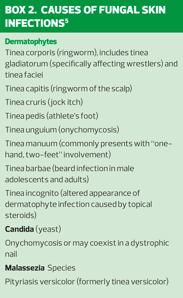
DERMATOPHYTE INFECTIONS
Dermatophytes are fungi that require keratin for growth. These fungi can cause superficial infections (tinea) of the skin, hair, and nails. Dermatophytes are spread by direct contact from other people (anthropophilic organisms), animals (zoophilic organisms), and soil (geophilic organisms), as well as indirectly from inanimate objects including upholstery, hairbrushes, hats.4 Dermatophytoses are known as tinea infections, which are further classified by the region of the body infected (Box 2).5 The classic appearance of a cutaneous tinea infection is a central clearing surrounded by an active border of redness and scaling, which gives rise to the more common name, ringworm, although a worm is not responsible and patients need reassurance that this is not the case. Tinea infections do not involve mucosa dermatophytes and only involve keratinized tissue. Tinea infections may be similar in appearance to many other skin conditions and are often misdiagnosed and, therefore, mistreated.2 There are three genera of dermatophytes – Trichophyton (T), Microsporum (M) and Epidermophyton (E). The most common organism in the UK is Trichophyton rubrum, except on the scalp where Trichophyton tonsurans and Microsporum canis predominate.6
Tinea Capitis – ‘Ringworm’ of the scalp
Tinea capitis (Figure 1) is the most common dermatophytosis in children but it can affect any age. In the UK a significant rise in the incidence and prevalence of cases of infection due to Trichophyton tonsurans has been seen. The main focus of this epidemic has been cities where there are either long-standing or more recently established Afro-Caribbean communities, although it is clear that infection can occur in any child irrespective of their ethnic origin. Another common organism causing tinea capitis is Microsporum canis.7
Transmission is by poor hygiene and overcrowding, and can occur through contaminated hats, brushes, pillowcases, and other inanimate objects.4 The main clinical characteristic of tinea capitis is hair loss, which is often accompanied by scaling. There may be symptoms such as itching and, more rarely, pain; often infections are asymptomatic. Hair loss may develop in single or multiple patches, but in addition individual hairs or small hair clusters may be involved. Hairs may be broken either above or at scalp level; where swollen broken hair stubs are prominent the pattern is known as black dot ringworm. Scaling may occur either in the presence or absence of hair loss and signs of inflammation such as erythema, pustule formation and crusting may be evident in very inflamed lesions. These inflamed lesions may result in a ‘kerion’ which is a boggy, sterile, inflammatory scalp mass with pustules and is commonly associated with zoophilic infections (animal host), but can also occur with anthropophilic ringworm (human host).6 Kerion formation is due to an immune response to the fungus.2 Cervical and occipital lymphadenopathy may be present.4 (http://www.pcds.org.uk/clinical-guidance/tinea-capitis-scalp)
Tinea faciei (face)
Tinea faciei resembles tinea corporis (ringworm). It may be acute (sudden onset and rapid spread) or chronic (slow extension of a mild, barely inflamed, rash). There are round or oval red scaly patches, often less red and scaly in the middle or healed in the middle. It is frequently aggravated by sun exposure. It may also present as a kerion (fungal abscess).8 (http://www.pcds.org.uk/clinical-guidance/tinea-faciei-face-and-barbae-beard)
Tinea corporis (body and limbs)
Tinea corporis (trunk, legs, and arms but excludes feet, hands and groin) affects all ages and is more common in males.8 It typically appears as single or multiple, annular, scaly lesions with central clearing, a slightly elevated, reddened edge, and sharp margination (abrupt transition from abnormal to normal skin) on the trunk, extremities, or face. The border of the lesion may contain pustules or follicular papules. The degree of itching is variable.4 The feet can act as the source of infection and should be examined and treated if they are involved.8 (http://www.pcds.org.uk/clinical-guidance/tinea-corporis-body-cruris-groin-and-incognito-steroid-exacerbated)
Tinea cruris – ‘jock itch’
Is more common in men than in women, occurs when ambient temperature and humidity are high and is frequently associated with tinea pedis. Occlusion from wet or tight-fitting clothing provides an optimal environment for infection. It affects the proximal medial thighs and may extend to the buttocks and abdomen. The scrotum tends to be spared and patients frequently complain of burning and pruritus. Pustules and vesicles at the active edge of the infected area, along with maceration, are present on a background of red, scaling lesions with raised borders.4 (http://www.pcds.org.uk/clinical-guidance/tinea-corporis-body-cruris-groin-and-incognito-steroid-exacerbated)
Tinea pedis – ‘athlete’s foot’
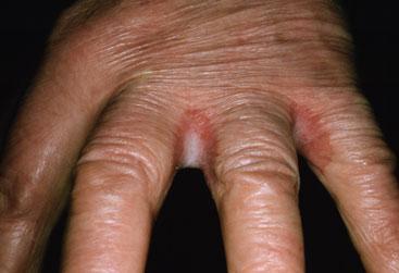
There are three common presentations of tinea pedis (Figure 2). Fissuring, maceration, and scaling in the interdigital spaces of the fourth and fifth toes with itching and burning characterise the common interdigital form. Another form has a moccasin- like distribution pattern in which the plantar skin becomes chronically scaly and thickened, with hyperkeratosis and erythema of the soles, heels, and sides of the feet and is usually caused by Trichophyton rubrum. A vesiculobullous form is characterised by the development of vesicles, pustules, and bullae usually on the soles.4 (http://www.pcds.org.uk/clinical-guidance/tinea-pedis-feet-manuum-hands-and-unguium-nails)
Tinea unguium – onychomycosis
A chronic progressive infection of the nail caused commonly by dermatophytes and less often by candida sp and moulds (Figure 3). One or just a few nail plates may be affected initially, but as time goes by there is a tendency for more to become involved. Toenails are involved more commonly than fingernails. The nails become thickened and discoloured, classically producing a yellowish-brown appearance, with cracking; the nail is more friable and raised by underlying hyperkeratotic debris in the nail bed.1,10 (http://www.pcds.org.uk/clinical-guidance/tinea-pedis-feet-manuum-hands-and-unguium-nails)
Tinea manuum (hands)
Usually affects one hand or, occasionally, both hands – but commonly presents with ‘one-hand, two-feet’ involvement, as it often occurs in patients with tinea pedis through scratching. The palmar surface is diffusely dry and hyperkeratotic. When the fingernails are involved, vesicles and scant scaling may be present.4 (http://www.pcds.org.uk/clinical-guidance/tinea-pedis-feet-manuum-hands-and-unguium-nails)
Tinea barbae (beard infection in male adolescents and adults)
Affects the beard and moustache area and closely resembles tinea capitis. There may be scaling, circular eryhtematous patches with broken surface hairs. There may be papules or pustules with exudation, crusting and the formation of a kerion.1 (http://www.pcds.org.uk/clinical-guidance/tinea-corporis-body-cruris-groin-and-incognito-steroid-exacerbated)
Tinea incognito (altered appearance of dermatophyte infection caused by topical steroids)
Occurs when a dermatophyte infection is modified because of treatment with a corticosteroid. Margins may be lost, and the area may be more widespread. Tinea incognito requires a thorough patient history and should be considered when a corticosteroid has been used to treat a rash that appeared to have cleared, but returned unresolved.2 (http://www.pcds.org.uk/clinical-guidance/tinea-corporis-body-cruris-groin-and-incognito-steroid-exacerbated)
CANDIDAL INFECTIONS OF THE SKIN (CANDIDIASIS)
Candida is part of normal body flora, but it is also a common cause of yeast infections. When the normal balance of flora is disturbed, an acute infection may occur and often presents as red lesions with accompanying satellite papules and pustules. Risk factors include antibiotics, corticosteroids, diabetes, obesity, immunosuppression, and immunodeficiency. Candida also thrives in warm, moist conditions.2 Common areas of infection include the mouth, genital region and intertriginous areas, finger or toe-webs (Figure 4), intra-mammary folds, axilla and groin and nails.3 Intertrigo can affect males and females of any age and is more common in overweight or obese patients, diabetic patients, the very young and very old due to reduced immunity, mobility and incontinence and hyperhidrosis (excessive sweating). Environmental and genetic factors also play a part and there may be an inflammatory or infectious origin with often an overlap between the two.11 Addressing the underlying predisposing risk factors is important especially in relation to intertrigo. Keeping the areas free from moisture and as dry as possible is a priority especially in the older person who may need help with hygiene needs.
MALASSEZIA INFECTIONS (PITYRIASIS VERSICOLOR)
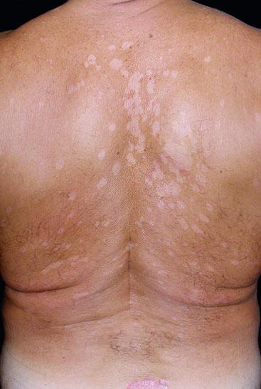
Malassezia yeasts (Malassezia furfur) were previously known as Pityrosporum yeast and are skin surface commensals which are associated with certain skin conditions, notably pityriasis versicolor (Figure 5). The term ‘pityriasis’ is used to describe skin conditions in which the scale appears similar to bran. The multiple colours arising in the disorder give rise to the second part of the name, ‘versicolor’. It sometimes called ‘tinea versicolor’, although the term ‘tinea’ should strictly refer to infection with a dermatophyte fungus. Pityriasis versicolor is a chronic usually asymptomatic scaling disorder which presents with macules, sharply marginated, round or oval in shape and varying in size on the upper trunk, upper arms and neck. In pale skin, the patches appear to be slightly dirty brown in colour, while in a darker skin or a skin that has been exposed to sunlight, the areas are hypopigmented.12 (http://www.pcds.org.uk/clinical-guidance/pityriasis-versicolor)
MANAGEMENT PRINCIPLES
Proper identification and treatment of fungal skin infections is paramount (Table 1). Good skin care, including regular bathing and complete drying of the skin, is essential for preventing fungal skin infections. Prolonged or frequent exposure to damp conditions and moisture should be avoided. Patients should be provided with a clear treatment plan and information regarding their condition. Objects that can transmit fungal spores (for example hats, combs, pillows, blankets, and scissors) should be discarded or disinfected (with bleach), where possible, to prevent re-infection or transmission of infection to others.13 Patients should not share towels, and towels, clothing, bedding should be washed frequently. If the infection affects the feet, patients should wear footwear that keeps the feet cool and dry, wear cotton socks and change to a different pair of shoes every 2–3 days. To reduce the risk of transmission they should be advised to avoid scratching affected skin, as this may spread the infection to other sites and should also avoid going barefoot in public places (they should wear protective footwear, such as flip-flops, in communal changing areas).14–16 Referral should be considered if:13
- The diagnosis is uncertain.
- There is no response to primary care management.
- Infection is severe or extensive.
- Infection is recurrent.
- The person is immunocompromised.
REFERENCES
1. Fitzpatrick TB, Johnson RA, Wolff K, et al. Color Atlas & Synopsis of Clinical Dermatology. 2001; New York: McGraw-Hill
2. Clinard VB, Smith JD. Cutaneous Fungal Infections. US Pharmacist. 2015; 40(4): 35-39.
3. Lawton S. Skin and Fungal Nail Infections. Independent Nurse Supplement. 2009;1:4-7
4. Hainer B. Dermatophyte Infections. Am Fam Phys 2003; 67:1: 101-108
5. ElyJ W, Rosenfeld S, Seabury Stone M. Diagnosis and Management of Tinea Infections. Am Fam Phys 2014;90:10:702-711.
6. Primary Care Dermatology Society (PCDS) Tinea – overview, 2016 http://www.pcds.org.uk/clinical-guidance/tinea
7. Primary Care Dermatology Society (PCDS) Tinea Capitis, 2016. http://www.pcds.org.uk/clinical-guidance/tinea-capitis-scalp
8. DermNetNZ. Tinea Faciei, 2003 http://www.dermnetnz.org/topics/tinea-faciei
9. Primary Care Dermatology Society (PCDS). Tinea corporis (body), cruris (groin) and incognito (steroid exacerbated), 2017. http://www.pcds.org.uk/clinical-guidance/tinea-corporis-body-cruris-groin-and-incognito-steroid-exacerbate
10. Graham-Brown R, Bourke JF. Mosby’s Color Atlas and Text of Dermatology. 1998; London: Mosby.
11. Lawton S. An overview of intertrigo: rashes affecting skin folds. Nursing and Residential Care 2017;19:1:30-33
12. DermNetNZ. Pityriaris versicolour, 2014. http://www.dermnetnz.org/topics/pityriasis-versicolor
13. NICE CKS. Fungal skin infection – scalp, 2014. https://cks.nice.org.uk/fungal-skin-infection-scalp#!scenario
14. NICE CKS. Fungal skin infection - body and groin, 2014 https://cks.nice.org.uk/fungal-skin-infection-body-and-groin#!scenariorecommendation:3
15. NICE CKS. Fungal Skin Infection – foot, 2014. https://cks.nice.org.uk/fungal-skin-infection-foot#!scenario
16. NICE CKS. Fungal Nail Infections, 2014. https://cks.nice.org.uk/fungal-nail-infection#!scenario


Related articles
View all Articles