Understanding blood results: the full blood count
Margaret Perry
Margaret Perry
RGN BSc(Hons) MSC
Advanced Nurse Practitioner, Linkway Medical Practice, West Bromwich
Practice nurses are often asked to take blood samples for a range of tests, including FBC. But what does it all mean?
The full blood count is a frequently requested test and is a useful tool in helping to diagnose a number of conditions. There are several components which make up the full blood count and each of these blood cells has a role to play in keeping the body healthy. Practice nurses are also often involved in reviewing test results and this article aims to help in their understanding of some of the parameters and make this task a little less daunting.
FUNCTIONS OF THE BLOOD
Blood is a type of tissue that is suspended in a watery substance known as plasma. The major functions of blood are:
- Transport of oxygen and carbon dioxide and food molecules such as glucose, amino acids and lipids
- Transport of sodium, calcium and bicarbonate
- Removal of waste products
- Transport of hormones
- Defending the body against invasion by foreign bodies and infections
About 95% of the body's blood cells are produced by the bone marrow, while other organs (the lymph nodes, liver and spleen) help regulate the production, destruction, and differentiation (developing a specific function) of cells.1
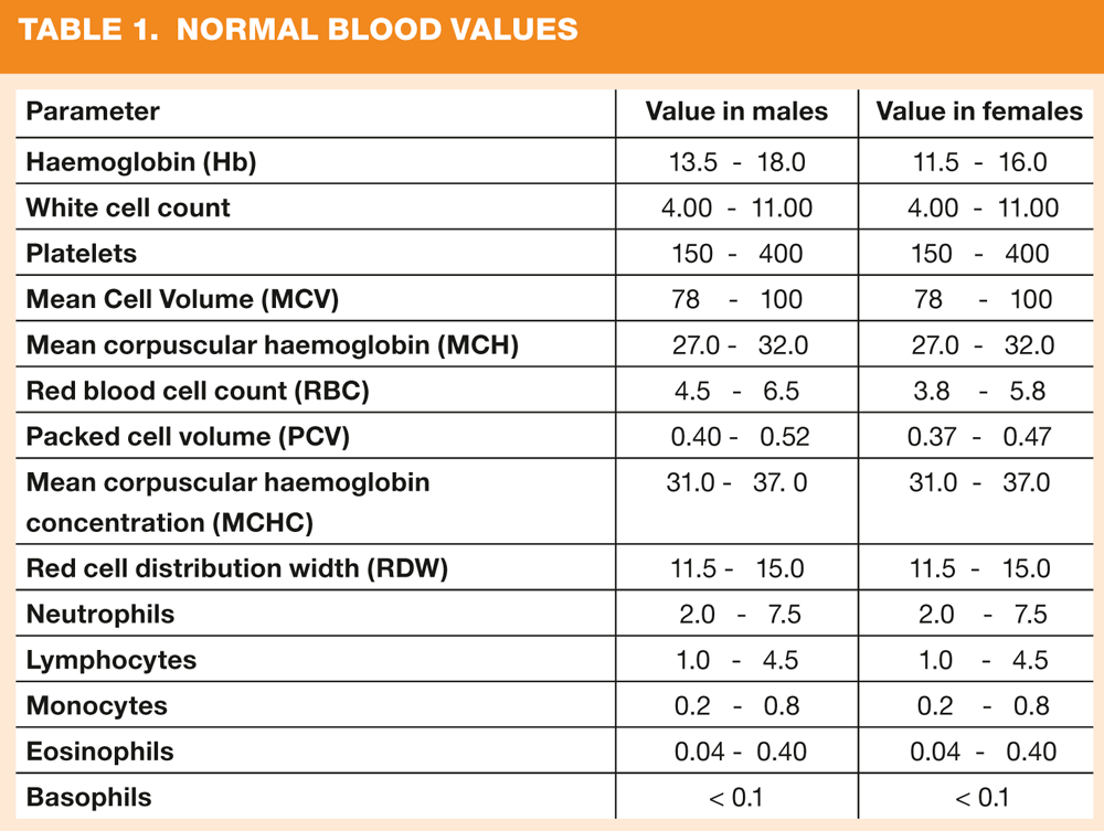
MEASUREMENTS AND NORMAL VALUES
Reference ranges vary slightly between males and females (Table 1 shows the normal values). The normal range describes the range where 95% of the normal healthy population will lie; the remaining 5% will fall outside the normal range, however they too are normal.2 It is important to remember that normal ranges are only a guide and there may be instances where parameters are well outside of this range but are considered normal for that person. One example of this is following splenectomy, where patients are often found to have a raised lymphocyte count but this is normal in the light of their medical history.
WHEN IS IT NEEDED?
A full blood count is often requested when the patient presents with symptoms, and the clinician is hoping to find clues to aid diagnosis when the result becomes available. There are however a number of conditions which will result in either increased or decreased values and in some cases will resolve spontaneously without any further treatment.
WHAT DOES IT MEAN?
Haemoglobin
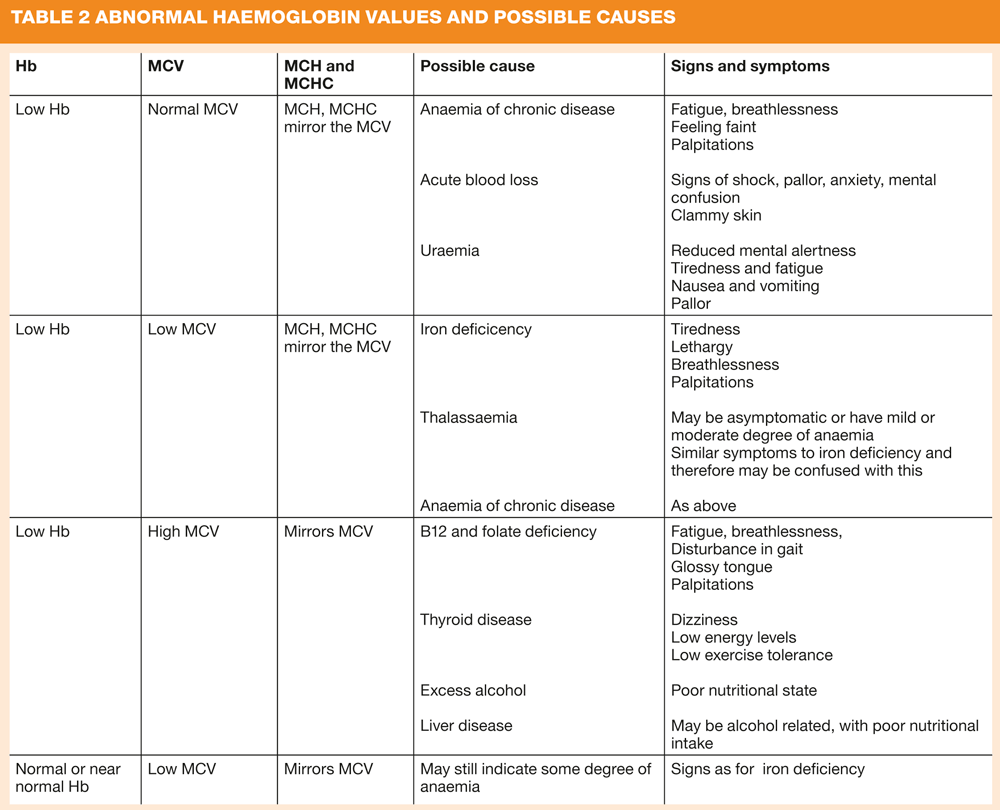
Measured in grams per decilitre (g/dl), haemoglobin (Hb) is the oxygen carrying protein and is part of the red blood cell. Its primary function is to deliver oxygen from the lungs to the tissues of the body and take carbon dioxide back to the lungs so that if can be exhaled. There are many reasons for low haemoglobin levels, and there may be instances where the level is slightly below target, is causing no symptoms and therefore no treatment will be needed. A low value is indicative of anaemia and should be considered alongside the mean cell volume (MCV) and mean corpuscular Hb (MCH) to aid diagnosis of the type of anaemia. Similarly there are numerous reasons for elevated haemoglobin levels, although some of the most frequent causes include COPD, emphysema, and heart failure.3 Suggested causes of abnormal Hb, MCV and MCH values are shown in Table 2.
Mean cell volume (MCV)
MCV is a measurement of the average size of the red blood cells. A high MCV indicates large cells, (macrocytic) a low MCV suggests small red blood cells. Abnormal values, either high or low help point towards the type of anaemia (see table 2).
Mean corpuscular haemoglobin (MCH)
MCH is considered one of the most diagnostically useful parameters,4 and is used to calculate the average amount of haemoglobin available to carry oxygen inside the red blood cell. The MCH mimics the MCV, therefore usually when MCV is raised so is MCH, and when MCV is low so is the MCH. However, beware that in elderly patients, the existence of comorbid conditions can affect the MCV and disguise the nature of the anaemia making diagnosis more difficult.4
Mean corpuscular haemoglobin concentration (MCHC)
This calculates the average concentration of haemoglobin inside red blood cells. It is low in certain types of anaemia and is high in conditions where the haemoglobin is abnormally concentrated. Decreased MCHC values (hypochromia) are seen in conditions where the haemoglobin is abnormally diluted inside the red cells, such as in iron deficiency anaemia, or thalassaemia. Increased MCHC values (hyperchromia) are seen in conditions where the haemoglobin is abnormally concentrated inside the red cells, such as in hereditary or autoimmune spherocytosis, a condition where red blood cell survival is decreased because of a structural defect in the red cell membrane.5
Packed cell volume
The packed cell volume (sometimes referred to as the haematocrit) is a measurement of the percentage of blood made up of cells. The value is reduced in conditions such as anaemia, leukaemia, bone marrow failure and multiple myeloma. It is increased in conditions causing dehydration which may occur as a result of diarrhoea or severe burns.
Red cell distribution width (RDW)
This is a measure of the size and shape of red cells. The value is increased in certain anaemias where variation in cell size and shape occurs. Red cell distribution width has been shown to be an independent predictor of mortality in patients with heart failure and coronary heart disease.6 Patients with haemoglobin levels above 10.4 and a high red cell distribution width have a significantly increased mortality risk.
Red cell count
This measures the number of red cells per litres of blood. Abnormally low values may indicate anaemia occurring as a result of iron deficiency or blood loss, bone marrow failure or abnormal destruction of red blood cells. Abnormally high values may be found in lung conditions such as chronic obstructive pulmonary disease (COPD), dehydration and kidney disease.
Platelets
Platelets are measured in thousands per cubic millimetre of blood and they are the smallest blood cell. They are important in forming blood clots, which they do by swelling and clumping together to form a plug at the site of bleeding. Too few platelets may lead to uncontrolled bleeding, whilst too high platelet counts may lead to formation of blood clots inside blood vessels and possible deep vein thrombosis or embolism. Platelet size is of diagnostic significance, particularly if considered in relation to the platelet count.7 Small or normal-sized platelets in association with thrombocytopenia suggest that the cause is a failure of bone marrow production, whereas thrombocytopenia with large platelets is more likely to be caused by peripheral destruction or consumption of platelets with the bone marrow responding by increasing platelet production.7
White cell count
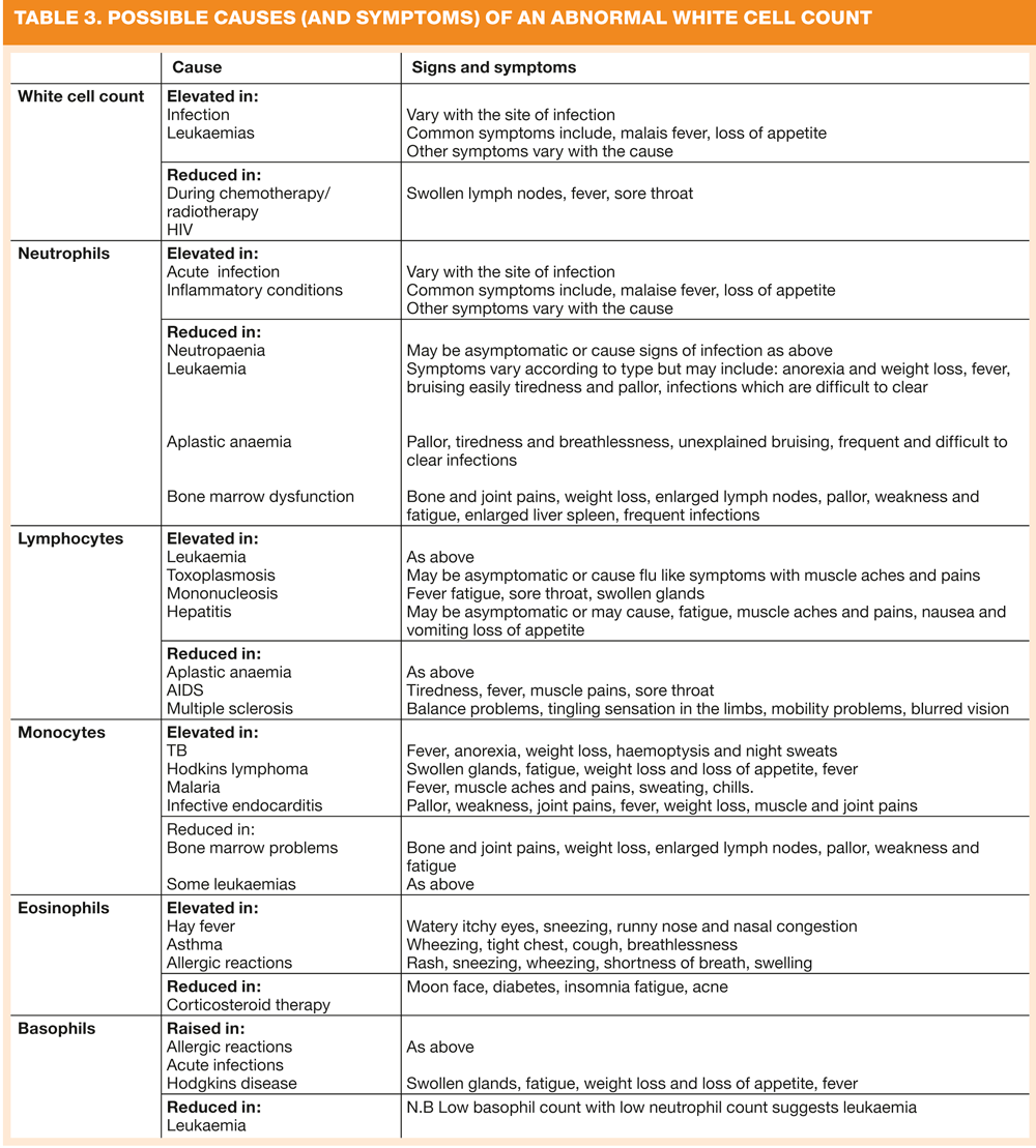
This measures the total number of white blood cells (leucocytes) but to give any meaning it should be viewed alongside the white cell differentials shown below. Each of these parameters has role to play in keeping the body healthy. Table 3 shows of the causes of elevated and reduced white cell count and white cell differentials.
Neutrophils are the most numerous of the white cells and they are active in the removal of invading bacteria or fungal antigens.
Lymphocytes make up approximately 25% of the total white blood cell count although this can vary.8 Lymphocytes exist in two forms, B cells and T cells. B cells produce antibodies and T cells are involved in the recognition of foreign bodies and co-ordinating the immune response.
Monocytes make up approximately 5-10 per cent of the total white cell count and function in the ingestion of bacteria and other foreign particles.8
Eosinophils are similar in size to basophils and are important in allergic reactions and parasaitic infections. They participate in engulfing and killing bacteria and other microorganisms and accumulate in excess numbers as part of a reaction to antigenic stimulations.9
Basophils are a type of cell that contains large amounts of histamine, which plays an active role in the allergic response.
P blood film
A P blood film (also called a peripheral blood film or blood smear) may be requested by the clinician or undertaken by the laboratory when components of the full blood count and/or white cell differential count are abnormal. This further test is useful in helping to identify cells which are poorly developed or abnormal in appearance. A blood smear can have an important part in the speedy diagnosis of some infections or in the differential diagnosis of anaemia and thrombocytopenia and in the identification and characterization of leukaemia and lymphoma.7
Putting the picture together
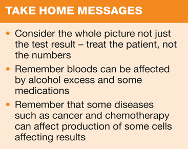
The patient's presenting symptoms and history play a vital part in pointing to a diagnosis. However, the full blood count result can often elicit abnormalities which were not considered, and on some occasions may identify a need for further investigation. Alternatively the result may simply confirm the clinician's suspicions. Hopefully a better understanding of the parameters which make up the full blood count will make the task of reviewing results a little easier.
REFERENCES
1. University of Maryland Medical Centre (2008) Blood diseases: Facts about blood http://www.umm.edu/blood/blood.htm
2. O' Sullivan 2010 The Full Blood Count. The Leeds Teaching Hospital NHS Trust http://www.pathology.leedsth.nhs.uk/pathology/ClinicalInfo/CommonTestsampInvestigations/FullBloodCount.aspx
3. Mayo Foundation for Medical Education and Research (2010) High haemoglobin count http://www.mayoclinic.com/health/high-hemoglobin-count/MY00112/DSECTION=causes
4. Bewick K and Metcalf P (2011) Anaemia. Geriatric Medicine 41 (8) 399-301
5. American Association for Clinical Chemistry (2008) Complete blood count. http://labtestsonline.org/understanding/analytes/cbc/tab/test
6. Poludasa S., J.D, Weedon J., Khan W and Cavusoglu E (2009) Red cell distribution width as a predictor long term mortality in patients undergoing percutaneous coronary intervention Journal of Thrombosis and Haemostasis 102 (30 581-7
7. Bain (B) 2003. A beginners guide to Blood cells. Ch3 Assessing white cells and platelets. E books http://www.bloodmed.com/home/ebook.asp?book=0865427178&id=3
8. American Association for Clinical Chemistry (2011) Lab test online: Complete blood count http://labtestsonline.org/understanding/analytes/cbc/tab/test
9. Collins M. (2004) What is an eosinophil? Cincinnati Children's Hospital and Medical Center http://www.apfed.org/downloads/What_is_an_Eosinophil.pdf
10. Bain B (2005) Diagnosis from the Blood Smear New England Journal of Medicine. 353:498-507
http://www.labtestsonline.org.uk/understanding/analytes/cbc/test.html


Related articles
View all Articles