Malignancies
Dr Mike Wyndhamis a GP in a suburban practice in Edgware MiddlesexHe has always been a keen photogr...
Dr Mike Wyndhamis a GP in a suburban practice in Edgware MiddlesexHe has always been a keen photographer, and has been taking medical photographs for more than 25 years. He has been a regular contributor to the GP press and his work has also appeared in the British Medical Journal, The Pharmaceutical Journal and various medical text books
Malignancy may present with very obvious signs such as haemoptyis, rectal bleeding or haematuria. However, it may present more subtly with nail changes such as clubbing. Cancer may metastasise to other organs of the body, and the skin may be affected too, with somewhat atypical looking lesions. Direct invasion through the skin from either primary or secondary lesions may present practical nursing difficulties, as can be seen in some of these images.
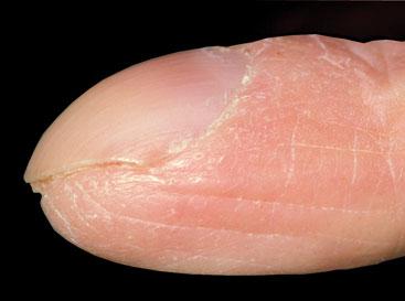
CLUBBING
Clubbing is a bulbous change of the end of the finger which will cause a change in the angle between nail fold and nail plate which will amount to greater than 180 degrees. It is usually painless. It may be bilateral but sometimes unilateral. There are many causes of clubbing which may range from cyanotic congenital heart disease to inflammatory bowel disease, pulmonary disease such as pulmonary fibrosis, empyema, sarcoidosis. Malignancies that may induce the condition, include lung cancer, Hodgkin's disease and thyroid cancer.
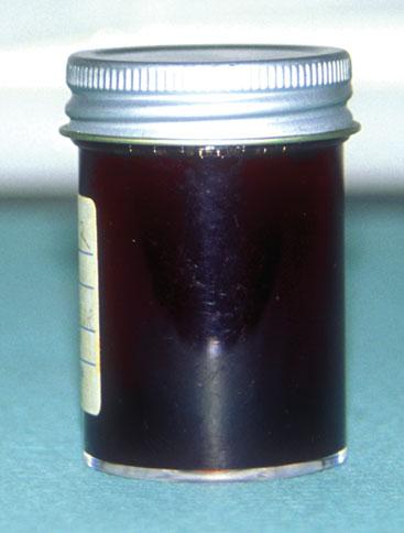
HAEMATURIA
There are many causes for macroscopic blood in the urine and it is a symptom that needs to be taken seriously, particularly if it is painless. Urinary tract infections may cause the urine to turn pink (diluted blood) and will be associated with dysuria. However, I have never encountered (in my 35 years' experience) a UTI to cause haematuria as shown in the picture. It should not be forgotten that beetroot and rifampicin may cause a reddish colouration to the urine so it is essential that a dipstick of the urine is carried out to confirm the presence of blood. Causes include renal stone, glomerulonephritis and tumours of the urinary tract. Anecdotally, a urologist colleague told me, one of the commonest causes of haematuria is aspirin when taken by people who have had prostatic surgery.
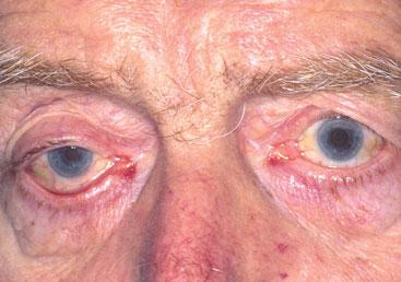
HORNER SYNDROME
Horner syndrome manifests as a triad of partial ptosis (drooping of the eyelid), constricted pupil (miosis) and loss of sweating in that half of the face. This results from a problem with the sympathetic nerve supply to that area. Causes include damage to the brachial plexus, brainstem stroke, migraine, and dissection of the internal carotid artery. The syndrome may also arise from problems affecting the apex of the lung, such as infection and Pancoast's syndrome. The latter is caused by a lung cancer in this region, which may be associated with severe pain in the shoulder region and compression of the local vasculature.
BREAST CANCER
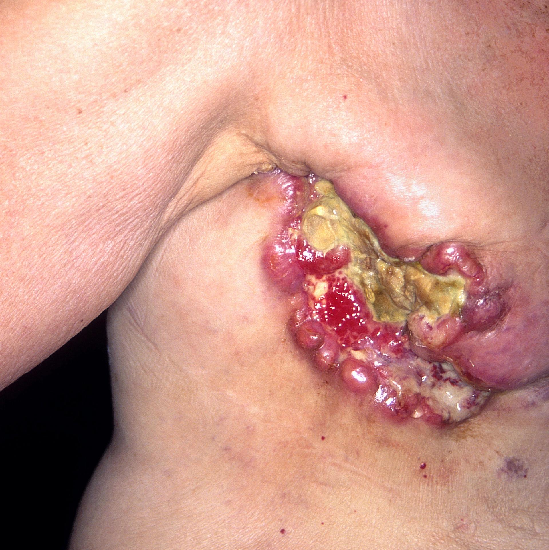
The diagnosis of cancer often creates fears about the likely impact of treatment or the uncertainties of long-term survival. Depression is common. Mastectomy may be considered disfiguring, and may lead to psychosexual problems. Women may also be worried by practical problems such as what to wear following a mastectomy, Advice and breast reconstruction may overcome some of these issues. But might these factors also prevent a women from asking for help, thus allowing a cancer to grow insidiously? Even the toughest practitioner would find the appearance of this fungating breast cancer very difficult to cope with. The patient was adamant that she did not want any palliative surgery.

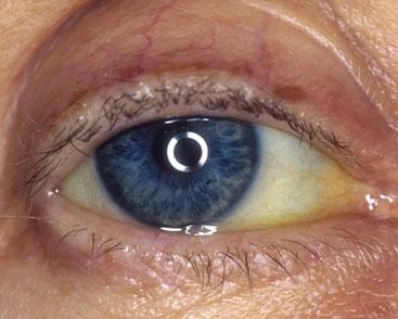
JAUNDICE
Jaundice starts to become visible when the serum bilirubin is above 35 micromols/litre. Jaundice may be sub-divided into pre-hepatic, hepatic and post- hepatic. The commonest cause of pre-hepatic jaundice in general practice is Gilbert's syndrome, with an isolated rise in bilirubin and no evidence of haemolysis. Cirrhosis and infectious hepatitis will cause a hepatic jaundice, with a mild rise in alkaline phosphatase. Post-hepatic or obstructive jaundice will cause a marked rise in alkaline phosphatase, which may be caused by the presence of gallstones. Malignancies that may cause this problem are pancreatic cancer and cholangiocarcinoma
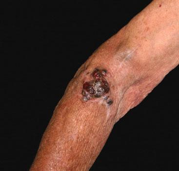
METASTATIC MELANOMA
The Breslow thickness is the best prognostic indicator for melanoma. It looks at the depth of invasion of the tumour measured in millimetres from the granular cell layer down to the last of the melanoma cells. If the thickness is <1mm then the 5 year survival rate is 95-100%. If it is > 4mm then the 5 year prognosis falls to 37-50%. This gentleman suffered a local recurrence following the original wide excision. A recurrence has developed below the original scar, with distal ulceration.