Nutrition and Bone Health in the UK: Calcium and vitamin D from cradle to grave
Dr Frankie Phillips
Dr Frankie Phillips
RD
Registered dietitian and Public Health Nutrition Consultant, Torquay, Devon
Diet is one of the key factors in forming healthy bones, and calcium, along with vitamin D, is essential for bone health. Too little vitamin D in utero and early childhood is leading to an increased incidence of rickets in the UK. Conversely, an ageing population has an increased risk of osteoporosis, but ensuring optimal nutrition for healthy bones is a lifelong aim, starting even before birth.
Bone is specialised connective tissue that is constantly changing. Osteoclast bone cells break down bone and osteoblasts form new bone tissue. If osteoclast activity exceeds osteoblast activity, bone mass is reduced, putting a patient at risk of fragility fracture. Osteoporosis is an extreme case of this bone remodelling.
Bone mass increases up to early adulthood, as large amounts of calcium (as well as some magnesium, zinc and phosphorus) are added to bone tissue. The skeleton grows, reaching peak bone mass (PBM) at around the age of 30-35 years. After this, bone mass starts to decline. Consequently it is important to strengthen bones early in life, although good nutrition is essential throughout life to maintain bone health, and offset the effects of ageing and bone loss.
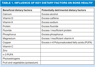
While genetic factors are the most important predictors of bone strength, accounting for 80% of variability in PBM, 20% of PBM can be accounted for by lifestyle choices, both dietary and non-dietary, to ensure optimal bone health. Non-dietary factors include physical activity, body weight, hormones, smoking and alcohol. Dietary factors are also significant, and a number of nutrients can potentially have a positive or negative impact on bone health (see Table 1). Adequate calcium can help to optimise PBM, but too much vitamin A (for example, from taking high dose cod-liver oil) can block the action of vitamin K, used to form the bone-hardening protein, osteocalcin, leading to osteopaenia. Nutrients may influence bone by various mechanisms, including alteration of bone structure, the rate of bone metabolism, the endocrine and/or paracrine system, and homeostasis of calcium and possibly of other bone-active nutrients.1
Probably the two most important nutrients for bone health are calcium and vitamin D. Vitamin D, amongst other roles, increases calcium absorption from foods.
Calcium is required for normal growth and development of the skeleton. Adequate calcium intake is critical to achieving optimal PBM and also modifies the rate of bone loss associated with ageing. In the UK, ideal calcium intake for adults is set at around 700mg per day. Requirements for calcium are higher during childhood, adolescence and lactation, but there is no increase in the recommended daily intake of calcium for older adults. The average woman consumes just over 700mg per day, the average man consumes just over 1000mg per day, thus for most adults, the body's requirements are met. Some concern has been raised over the high numbers of teenagers and young women with low calcium intake, and the implications this has on achieving peak bone mass and later bone health.2 Encouraging the inclusion of low-fat dairy products, such as semi-skimmed milk and low-fat yogurt can be a useful way of ensuring sufficient calcium in the diet.
Older men and women need to ensure that they include sufficient foods containing calcium (and vitamin D) to help protect their bones, especially since declining kidney function can impair vitamin D metabolism and calcium absorption. For older adults, low-fat dairy products can be a valuable source of calcium and skimmed milk powder can also be added to soups, sauces and custard to further fortify these foods. Oily fish, such as canned sardines and salmon can also help to increase calcium intake, especially if the soft bones are also eaten; oily fish additionally provides dietary vitamin D.
Women are particularly at risk of increased bone loss. From the menopause, women have declining levels of oestrogen. Since oestrogen helps the body to absorb calcium, low levels of oestrogen can increase risk of osteoporosis as the rate of bone loss, which starts in the mid-30s, accelerates. This is particularly true in the first years following menopause.
OSTEOPOROSIS IN THE UK
Osteoporosis is a debilitating disease that affects many older people. It is estimated that one in two women and one in five men over 50 years old will suffer an osteoporotic fracture,3 with immense implications for costs to the National Health Service, as well as the cost to individuals' quality of life.
As life expectancy increases, osteoporosis is likely to become an increasingly significant health problem, with fragility fractures having not only a significant effect on NHS costs, but also on personal costs in terms of quality of life. In the UK, it is estimated that the cost of treating all osteoporotic fractures in post-menopausal women will increase to more than £2.1 billion by 2020.3
Risk factors for osteoporosis
Bone mass in later life is largely dependent on PBM achieved during growth and the rate of subsequent age-related bone loss. Low bone mineral mass is the main factor underlying osteoporotic fracture, therefore achieving maximal bone mass during growth and reduction of loss of bone later in life are the two main strategies for preventing osteoporosis. Therefore, any factor that influences PBM or the loss of bone in middle-age will affect later osteoporosis risk.
Several factors are thought to influence bone mass. The interaction of genetic, hormonal, environmental and nutritional factors influences both the development of bone to peak bone mass at maturity and its subsequent loss. Some factors cannot be modified, such as gender, age, frame size, genetics and ethnicity; those that can be modified include hormonal status (especially sex and calciotropic hormone status), lifestyle factors including physical activity levels, smoking and alcohol consumption, and diet. In addition, the treatment of a variety of medical conditions may also influence bone health.
Glucocorticoids are widely used in the treatment of patients with chronic non-infectious inflammatory diseases, especially asthma, chronic obstructive pulmonary disease, rheumatoid arthritis and other connective tissue diseases, inflammatory bowel disease, and in organ transplantation. Although the beneficial antiinflammatory and immunosuppressive effects of glucocorticoids necessitate their use, adverse side effects are frequent. Osteoporosis and related fractures are among the most serious adverse effects; indeed, glucocorticoids are the most common cause of drug-related osteoporosis.4
Other drugs that can adversely affect bone density include;
- unfractionated heparin
- aromatase inhibitors
- gonadotropin-releasing hormone antagonists
- long-acting (depot) progesterone-containing contraception
- thyroid replacement therapy
- thiazolidinediones
- proton pump inhibitors
- selective serotonin re-uptake inhibitors
- antiepileptics, and
- calcineurin inhibitors.5
Before initiating prescriptions for any of these medications, patients' fracture risk should be assessed. When fracture risk is high consider medications with less skeletal impact. Medication use should also be regularly reviewed. In addition, calcium and vitamin D intake should be optimized.5
Supplements containing both vitamin D (which is usually listed as 'D3' or cholecalciferol) and calcium (as calcium carbonate, calcium citrate or calcium phosphate) may be recommended to individuals at risk of developing or with osteoporosis. Studies have shown that calcium alone cannot prevent or cure osteoporosis but having enough calcium is essential to maintaining bone strength and can play a vital role in preventing osteoporosis-related fractures, particularly in elderly women living in care homes who are more likely to have poor vitamin D levels.2
VITAMIN D
Vitamin D is not a 'true' vitamin, but describes a group of related pre-hormones, the two main forms of which are ergocalciferol (D2) and cholecalciferol (D3). Cholecalciferol is converted in the liver and kidneys to water-soluble calcitrol, which circulates as a hormone in the blood, regulating the concentration of calcium and phosphate and promoting healthy growth and remodelling of bone. Calcitrol has a crucial role in maximising absorption of calcium from the gut.
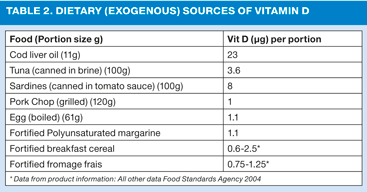
Unlike other vitamins, which are obtained from the diet (exogenous), vitamin D is found naturally in very few foods (see Table 2) and most vitamin D is produced from the action of sunlight on skin, not from diet. Endogenous synthesis of vitamin D occurs when skin is exposed to UVB radiation from sunlight. However, in latitudes above 40oN, synthesis of vitamin D occurs little if at all from October to April. Thus, during the winter months there is an increased reliance on dietary supply of vitamin D or supplements to boost stores of vitamin D, with some groups possibly being at risk of low vitamin D levels during the winter and early spring as stores are depleted, with potential adverse consequences for bone health. In particular, people who are housebound, who cover up their skin for cultural reasons, pregnant and breastfeeding women and their infants, and elderly people whose ability to synthesise vitamin D from sunlight is diminished, are more likely to have low vitamin D status.
VITAMIN D DEFICIENCY
A deficiency of vitamin D is characterised by inadequate or defective mineralization, or demineralisation, of the skeleton causing rickets in children and osteomalacia in adults. Concerns have been raised by the Department of Health that more attention is needed to ensure that pregnant and breastfeeding women and children under 5 years are advised to take a vitamin D supplement to prevent the rise of rickets in the UK.
Vitamin D deficiency is also more prevalent in patient with Crohn's disease, coeliac disease and cystic fibrosis.
Secondary hyperparathyroidism associated with low vitamin D status enhances mobilisation of calcium from the skeleton. Vitamin D deficiency is also an important contributor to osteoporosis through reduced intestinal absorption of calcium, increased bone loss, muscle weakness, and a weakened bone microstructure.1 A review of the evidence has shown that even marginal vitamin D deficiency is likely to have a significant negative impact on bone health: even if vitamin D deficiency is not sufficiently serious to cause rickets, it could prevent young adults from reaching their peak bone mass. In addition, studies have shown that poor vitamin D status during pregnancy has a detrimental effect on bone mineral content in the child, pointing to the need for vitamin D supplementation during pregnancy.1 Consequently it is thought that maintaining adequate vitamin D levels could help to achieve PBM and reduce risk of osteoporotic fracture.
RICKETS AND OSTEOMALACIA
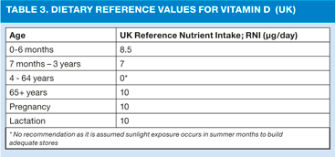
In the UK there is currently no recommended daily requirement for vitamin D for 4 - 65 year olds; it is thought that adequate vitamin D is produced with exposure to sunlight between April and September producing enough to last through the winter months.
However, concerns about the risks of skin cancer associated with sun exposure have resulted in more widespread use of high protection sunscreen products, and this is thought to be one of the underlying causes of the increase in prevalence of rickets that has been observed in recent years. Another reason for the increase may be the many hours that children spend indoors, playing computer games or watching television.
A further factor is that vitamin D deficiency is endemic in people with dark skin, especially South Asian, African Carribbean and Middle Eastern populations - prevalence in in otherwise healthy South Asian adults is as high as 94%. The prevalence of rickets in non-Caucasian children has been found to be 1.6%, and overall incidence is 7.5 per 100,000 children under 5 years.6
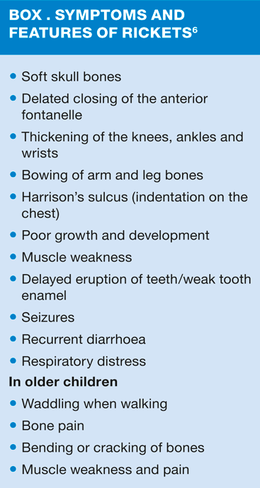
Symptoms and features of rickets in children and osteomalacia in adults include 'aches and pains' in bones, muscle weakness and sometimes visible enlargement of bones at joints e.g. at the wrists. (See Box 1). Fractures may occur with little or no recognised trauma. In adults, osteomalacia is increasingly identified after a finding of low bone density.
RECOMMENDATIONS
Current advice is that in fair skinned people, exposure of the face and arms to sunshine for about 20 to 30 minutes, twice or three times a week before applying a sunscreen should be enough to produce healthy levels of vitamin D in the summer in the UK, but people with pigmented skin would require longer or more frequent exposure to obtain the same vitamin D levels.7
For some age groups (small children and people over 65 years) and for pregnant and lactating women, specific recommendations for vitamin D requirements are set in the UK (see Table 3).
The Endocrine Society recommends screening for vitamin D deficiency in people at risk of deficiency, defined as 25(OH)D below 20ng/ml (50nmol/l), and that adults who are vitamin D deficient should be treated with 50,000 IU of vitamin D once a week for 8 weeks or its equivalent of 6000 IU daily.8 Full treatment recommendations are available at www.endo-society.org.
Although breastfeeding is recommended, prolonged breastfeeding without vitamin D supplementation can lead to low vitamin D status in the child, and to the development of rickets in infants and toddlers. Dietary intake of vitamin D is low for many people in the UK (around 2-4 ug per day), and vitamin supplements may be indicated as few of the foods rich in vitamin D are commonly eaten in sufficient amounts (see Table 3). For children under 5 and pregnant and breastfeeding women, Healthy Start vitamin drops and tablets are recommended. In some areas in the UK, Healthy Start vitamins for women (containing vitamin C, Vitamin D and folic acid) are provided free of charge for all pregnant women, as well as those in receipt of benefits; in other areas, they may be purchased.
CONCLUSION
Good bone health, and the prevention of both rickets and osteoporosis, and other bone conditions, depends on good nutrition through life, with calcium and vitamin D as the most important nutrients for strong healthy bones. Dietary factors can have a significant impact on bone mass and bone loss.
Dietary sources of vitamin D are limited and exposure to sunlight and vitamin D supplementation are important to prevent poor vitamin D status. In light of the high prevalence of low vitamin D status among population groups in the UK, strategies are needed to address this public health problem, with focus on adequate vitamin D intake from infancy through to old age.
REFERENCES
1. Cashman K. Diet, nutrition and bone health . The Journal of Nutrition 2007;137(11 Suppl):2507S-2512S.
2. Theobald HE. Dietary calcium and health. Nutrition Bulletin 2005;30:237-277.
3. National Osteoporosis Society. Facts and Figures. Available at: http://www.nos.org.uk/Document.Doc?id=47 4. American College of Rheumatology. Treatment of steroid induced osteoporosis; ACR task force on Osteoporosis Recommendations for the prevention and treatment of glucocorticoid-induced osteoporosis. Available at http://www.rheumatology.org/practice/clinical/guidelines/osteo.asp 5. Pitts CJ, Kearns AE. Update on medications with adverse skeletal effects. Mayo Clin Proc 2011;86:338-343
6. Lowden J. Rickets: concerns over the worldwide increase. J Fam Health Care. August 2011. Available at: http://www.jfhc.co.uk/Content/Doc.aspx?id=1563 7. Pearce SHS, Cheetham TD. Diagnosis and management of vitamin D deficiency. BMJ 2010;340:b5664
8. Endocrine Society Clinical Guidelines. Evaluation, treatment and prevention of vitamin D Deficiency. Available at: http://www.endo-society.org/guidelines/final/upload/final-standalone-vitamin-d-guideline.pdf
