Practical Skills in Primary Care Part 2
Stephanie Garner
Stephanie Garner
RGN, BSc Nurse Practitioner
MSt (Cantab) Primary and Community Care, Independent/Supplementary Prescriber
Advanced Nurse Practitioner, Wisbech, Cambridgeshire
In the second of our two-part series on the essential tasks routinely undertaken by practice nurses and healthcare assistants we focus on the practical clinical skills of venepuncture, blood pressure monitoring and taking an ECG
TAKING A BLOOD SAMPLE
Venepuncture, otherwise known as phlebotomy, is one of the most commonly performed invasive procedures.1 The process involves inserting a needle into a vein to take a representative sample of the circulating blood which is analysed for diagnostic, monitoring or screening purposes.
Preparation
Before starting the procedure collect everything you require. This will include relevant personal protective equipment (PPE) and infection control equipment together with the request form and appropriate venepuncture collecting system. There are various different collection systems available such as Vacutainer®, Monovette® or Vacuette®, so ensure you are familiar with the system used within your local area, remembering that the bottle lid colour (determining the presence or absence of preservative) differs between systems. All equipment should be sterile and single use only.
With any procedure involving body fluids particular attention should be paid to Infection Prevention and Control (IPaC). With regards to venepuncture this should include hand hygiene, aseptic technique, protective clothing and sharps disposal.
For the patient, venepuncture may be a frightening experience and this should be given adequate consideration during the preparation process.
Procedure
It is essential you confirm the patient's details using a minimum of two forms of identification, carefully checking the request form for which samples are required. Any lapse of concentration at this point could have disastrous effects.
Ensuring the patient's comfort, select an appropriate vein to take the sample from avoiding affected sides following cerebral vascular accidents (CVAs) or post mastectomy where lymphatic drainage may be affected. Also avoid sites where peripheral vascular devices or fistulas are in situ, or where bruising, infection, inflammation or scarring is present. You should also consider that taking a sample close to a site where intravenous fluids are being administered may affect the result.
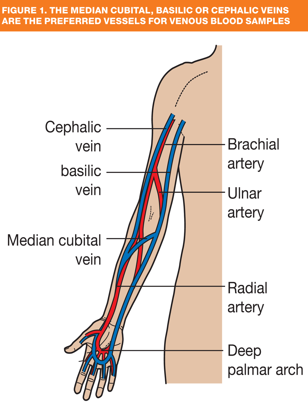
The most commonly — but not exclusively — used veins are those in antecubital fossa: the median cubital, basilic or cephalic veins. (Figure 1) These are preferred because they are easily accessible, a good size and cause less discomfort when punctured. A combination of visualisation and palpation should be used to determine vein choice as this ensures veins are selected rather than arteries or tendons, and enables valves to be avoided which could hinder flow.
A single use tourniquet should be applied to the upper arm 5—10cm above the proposed puncture site. It should be tight enough to impede the venous flow, but not occlude arterial flow (a radial pulse should still be present). You may also ask the patient to open and close their hand to encourage venous filling, thus dilating the vein.
There is conflicting advice regarding skin cleansing therefore you should follow local policy: if an alcohol preparation is used, it should be allowed to dry completely before taking the sample using the process described by the chosen system manufacturer.
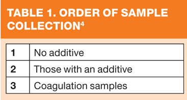
If multiple samples are required they should be collected in the order shown in Table 1 to avoid haemolysis.
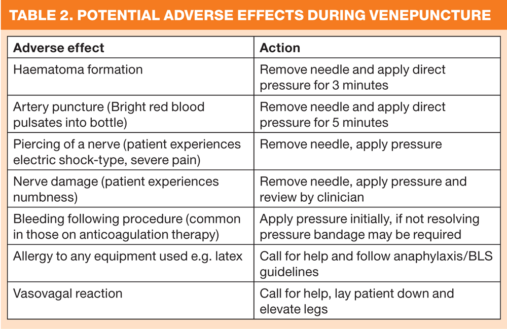
It is the clinician's responsibility to be able to deal with any adverse effects that may occur during the process (Table 2).
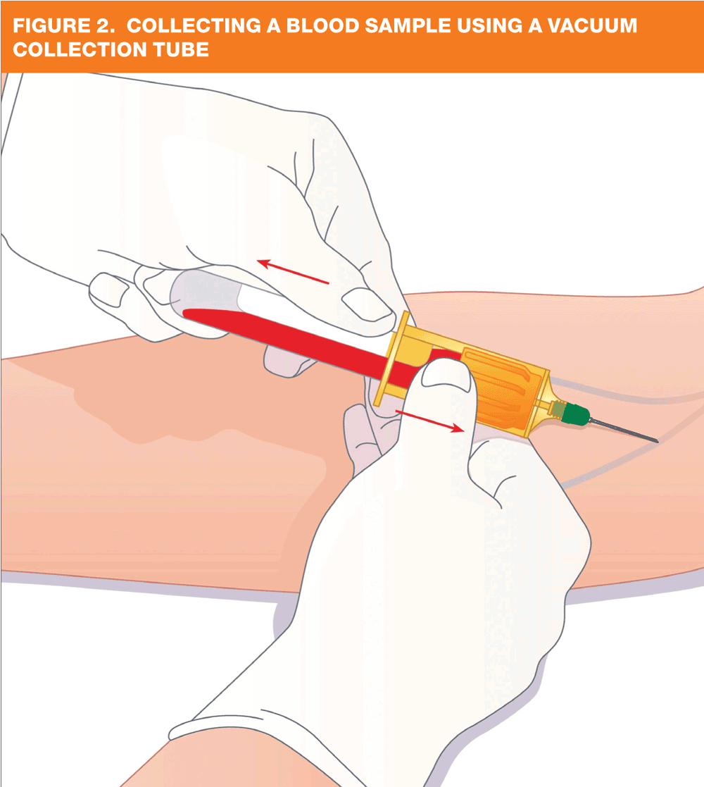
Once the last sample has been collected, release the tourniquet, remove the needle and place a swab over the site, applying pressure. Dispose of equipment, immediately label the samples and — as the gold standard procedure dictates — double check any cross matched samples.
Attend to the patient's comfort, applying a dressing if required and give relevant advice on post procedure care including when and how results will be available.
TAKING A BLOOD PRESSURE
Blood pressure (BP) measures the force that the heart uses to circulate blood around the body, the systolic (top) recording being when the pressure is at its highest and the diastolic at its lowest.5
BP may be used as an indicator of risk for other diseases such as coronary heart disease (CHD) or used to assess the severity of a patient's condition, as in hypovolaemic shock.
A patient with a persistently raised BP (hypertension) is at much greater risk of CHD especially when this is combined with other lifestyle risk factors and/or multiple diseases; hence it is important to monitor BP, either to make an initial diagnosis or to monitor the effect of any medication. The NICE clinical guideline on hypertension (CG127) sets out guidance on the regularity of monitoring and optimal target levels.6
Blood pressure measuring devices
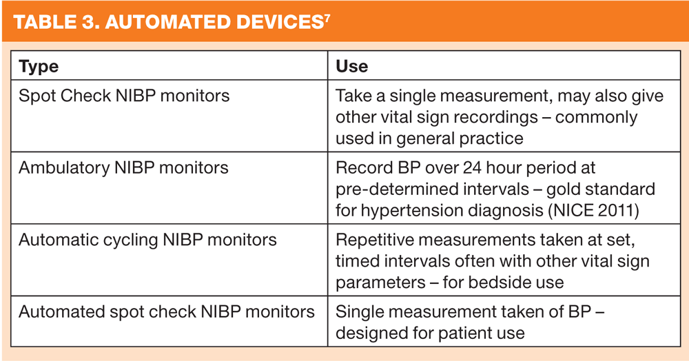
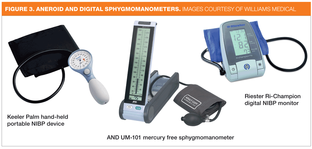
The gold standard for measuring BP is with the traditional non-invasive BP (NIBP) mercury sphygmomanometer using the auscultatory method (Korotkoff Sounds). However, with environmental concerns about mercury use and the subsequent restrictions on its use, NIBP aneroid gauge or electronic pressure transducer devices are more commonly used,7 despite concerns about their accuracy. Different types of device are shown in Figure 3. As all machines differ, you should familiarise yourself with the devices you will be using and to reduce the possibility of inaccuracy, ensure they are calibrated regularly, according to the manufacturer's instructions.
There are currently four types of automated device (see Table 3).
Appropriate cuff size
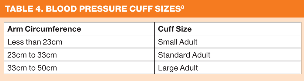
An appropriate cuff should be used to ensure accuracy of the recording. The upper arm should be measured at its widest circumference to determine the cuff size required (see Table 4).
Where possible the patient should not smoke, eat, drink caffeinated beverages or take alcohol in the 30 minutes prior to the reading and should empty their bladder if required. Tight clothing on the arm should be removed and the patient positioned comfortably ensuring the arm to be used is outstretched and at heart level.
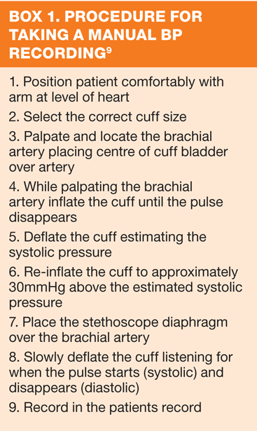
Apply the correct sized cuff, politely ask the patient not to speak during the procedure and either activate the device as per manufacturer's instructions or take the manual reading (Box 1). Two readings should always be taken with the lowest being recorded.
Automated devices
Automated devices may not be able to determine accurate BP in patients with irregular heart rates such as atrial fibrillation or in those with a regular heart beat of less than 50 beats/minute. Manual BP in these cases may be preferable.
TAKING A 12 LEAD ELECTROCARDIOGRAM
An electrocardiogram (ECG) is a test that records the electrical activity of the heart in wave form. It is performed either for monitoring or for diagnostic purposes, and it is essential that it is performed accurately.
Preparation
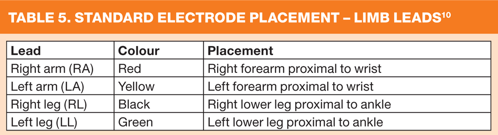
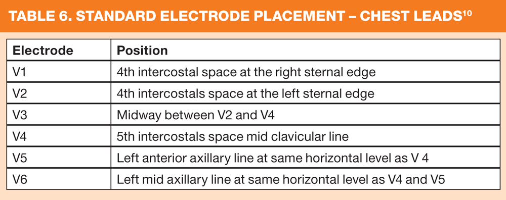
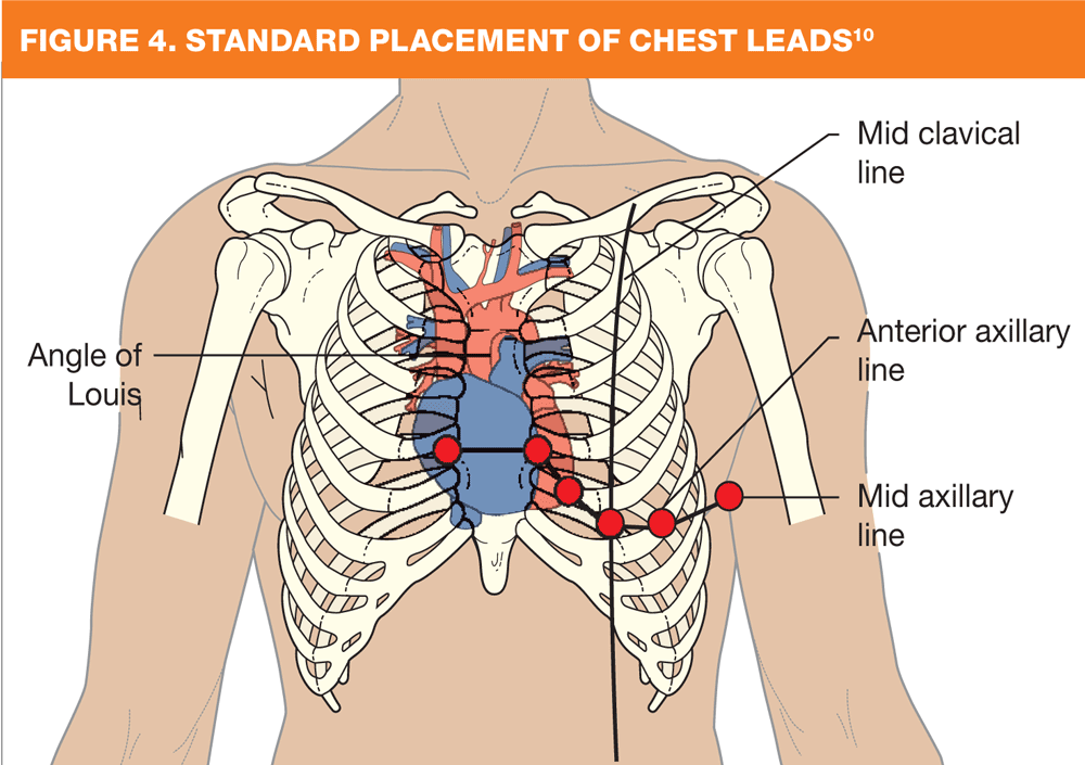
Maintaining privacy and dignity, the patient should be in a recumbent position that allows access to their chest, legs and arms so the electrodes can be placed accurately. Electrodes placement should be as per the Society for Cardiological Science and Technology recommendations as shown in Figure 4 and listed in Tables 5 and 6.10 On occasion, it may be appropriate to deviate from these standard placements, and if this is the case, it should be recorded on the ECG.
For accurate recording electrodes must have a good contact with the skin. Greasy body lotions, creams or perspiration, together with excess body hair can affect adhesion, and whenever possible, should be removed before the procedure.
Once the leads have been placed, ensure the patient is relaxed as any movements together with external electrical interference can affect the clarity of the tracing making interpretation more difficult.
The ECG should be labelled with the patient's details including the date and time the recording was taken together with any supporting information that may be relevant to why it was being undertaken.
CONCLUSION
It is important to maintain practical clinical skills to a high standard. Monitoring trends forms an important part of individualising patient care plans, allowing provision of the best quality care, and ensuring a good patient experience. Registered nurses should regularly review current evidence-based guidelines to make certain that not only are their skills being maintained so they continually work within the NMC Code,2 but also to make sure protocols and policies are kept up to date with the latest guidance. In this way, they will also ensure that other members of the team are able to share that knowledge and continually provide high standards of care.
REFERENCES
1. Gabriel J. Venepuncture and cannulation: considering the ageing vein. British Journal of Nursing. 2012;21(Suppl 1):22-28
2. Nursing & Midwifery Council. Delegation. Available at: http://www.nmc-uk.org/Nurses-and-midwives/advice-by-topic/a/advice/Delegation/
3. Nursing & Midwifery Council (2008) The code: Standards of conduct, performance and ethics for nurses and midwives. Nursing & Midwifery Council. London
4. Becan-McBride K. Laboratory sampling: does the process affect the
outcome? Journal of Intravenous Nursing 1999;22(3):137—142
5. British Heart Foundation (2013) Blood Pressure. Available http://www.bhf.org.uk/heart-health/conditions/high-blood-pressure.aspx
6. National Institute for Clinical Excellence (2011) Hypertension Clinical management of primary hypertension in adults CG127. NICE. London
7. MHRA (2013) Blood pressure measurement devices. Available: http://www.mhra.gov.uk/Publications/Safetyguidance/DeviceBulletins/CON2024245
8. British Hypertension Society (2006) Blood pressure measurement. Fact file. Available: http://www.bhsoc.org/files/5213/3363/9181/Factfile_2006_1_Blood_Pressure_Measurement.pdf
9. British Hypertension Society (2013) Blood pressure measurement with manual blood pressure monitors. Available: http://www.bhsoc.org/files/9013/4390/7747/BP_Measurement_Poster_-_Manual.pdf
10. The Society for Cardiological Science and Technology (2010) Clinical Guidelines by Consensus - Recording a 12 lead electrocardiogram - an approved methodology. Available at: http://www.scst.org.uk/resources/consensus_guideline_for_recording_a_12_lead_ecg_Rev_072010b.pdf
Related articles
View all Articles