Assessing chest pain in primary care
Beverley Bostock-Cox RGN, BSc, MScNurse Practitioner Sky Blue Medical Centre, Coventry. Clinical Le...
Beverley Bostock-Cox RGN, BSc, MScNurse Practitioner Sky Blue Medical Centre, Coventry. Clinical Lead, Education for Health, Warwick
It is increasingly common for nurses to provide first point of contact care for people with a range of signs, symptoms and conditions - including chest pain.
Nurses, in both primary and secondary care, are often called upon to provide first point of contact care for people with a range of signs, symptoms and conditions. In the case of chest pain, the importance of thorough history taking, careful assessment, including examination, and the use of objective tests to ascertain whether the pain is likely to be cardiac or non-cardiac in origin, cannot be overstated. Thorough assessment will allow the safest and most appropriate course of action to be taken.
THE IMPORTANCE OF CORRECT DIAGNOSIS
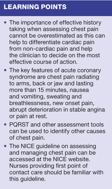
Chest pain is a common presenting complaint in general practice.1 Although most chest pain will turn out to be non-cardiac in origin once a full history has been taken and a thorough assessment carried out,2 it is imperative that due consideration is given to excluding acute and serious heart disease in each case. When a patient presents with chest pain the most important aspect of the initial assessment is to take a careful history.3 This will provide the core of information that will allow the clinician to decide whether the pain is likely to be Acute coronary syndrome (ACS), stable angina or non-cardiac chest pain, so that appropriate and, in some circumstances, life-saving action can be taken.
ACS is an umbrella term which covers myocardial infarction and unstable angina, both of which are potentially fatal.4 The maxim 'time is tissue' should be borne in mind - any delay in making a diagnosis of ACS can result in delay in initiating treatment and may result in permanent damage to the heart muscle, or even death.
Once acute cardiac causes for chest pain have been largely excluded, other possible diagnoses can be considered. Those patients who are more likely to have a serious cardiac or non-cardiac cause for their symptoms, can be referred for urgent care immediately. At the same time, those with less serious conditions can be treated appropriately without the need for inappropriate referrals, which may needlessly increase anxiety levels for patients and their families and waste limited NHS resources.5
In primary care settings a minority of chest pain presentations are due to acute cardiac causes, although in patients presenting in A&E departments the number due to ACS increases significantly. It is perhaps important to stress that, in cases of doubt, it is better that people are admitted unnecessarily than for cardiac pain to be misdiagnosed.
HISTORY TAKING
In the past, making a diagnosis was deemed to be the remit of doctors and the nurse's role was to provide appropriate, evidence-based care after the diagnosis had been made. Indeed, as recently as the past decade, when nurse prescribing was introduced, it was stated that the supplementary nurse prescriber could only prescribe for a condition once the diagnosis had been made by a doctor.6 Not all currently practising nurses will have been taught the history taking skills required for making an accurate diagnosis. That nurses are properly trained and equipped for their role is a fundamental part of the nursing code of conduct. Before taking on and accepting accountability for first point of contact care nurses require to be adequately trained and able to prove their competence.
There are a number of useful guides that explain how to take a good clinical history3,7 but, even when competence has been achieved, it is worthwhile taking time to practice history taking skills as often as possible.
Notes should be clear and unambiguous. The correct terminology when describing anatomical sites, symptoms and so on should always be used so that findings are not open to misinterpretation by another clinician. History taking should start with the presenting complaint and go on to cover:
- Past medical history
- Family history, if appropriate
- Medication history, including over the counter and recreational drugs
- Psychosocial elements, such as anxiety
- Employment status
- Relationship issues
- Smoking history and alcohol consumption.
All of these factors will help identify those who have risk factors for coronary heart disease (CHD) and those presenting with symptoms more likely to be non-cardiac, such as dyspepsia, lung disease, anxiety or medication side effects. Taking a full history can take some time and it is important to be systematic so that vital points (which a patient may not spontaneously reveal) are not missed.
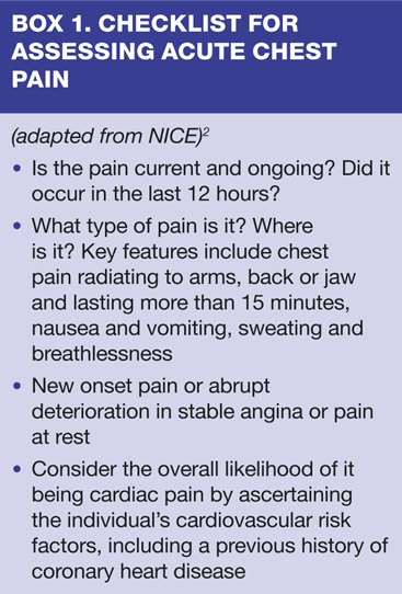
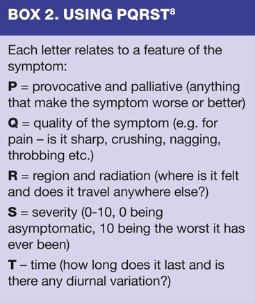
A checklist for assessing people presenting with acute chest pain is provided in Box 1. In addition there are several 'aide memoire' tools which can be used to help ascertain the main features associated with a symptom such as chest pain. These include PQRST8 (Box 2), SOCRATES9 and OLDCART.10
Using PQRST
Here, as an example, is an explanation of how to use the PQRST tool to help identify elements which increase or decrease the possibility of cardiac or non-cardiac pain and also to assess the impact of the pain on the individual. In a classic case of cardiac chest pain, PQRST may reveal some or all of the following typical features:
P - made worse by activity or emotion, relieved by rest or GTN
Q - pain is crushing, like a tight band or pressure around the chest
R - it is often central, retrosternal, radiating into the jaw, back, shoulders and/or arms
S - severity will vary but may be very severe
T - in stable angina it is it is short-lived; more severe and prolonged symptoms are more likely to be related to ACS and will require urgent admission and intervention.2
Compare this with chest pain caused by reflux oesophagitis...
P - made worse by lying flat, relieved by antacids or similar medication
Q - sharp pain
R - epigastric radiating into the gullet and/or throat
S - severity will vary but is more likely to be moderate
T - intermittent11
...or musculoskeletal pain:
P - made worse by specific movements or pressure on the affected site, relieved by changing position or by standard analgesia
Q - aching, maybe sharp if there is nerve involvement
R - specific to the site affected and can often be pointed to directly
S - from no pain in one position to severe in another
T - intermittent, short-lived or chronic.11
Completing the history
Other factors which need to be identified when taking a complete history of a patient presenting with acute chest pain include:
- Any previous or family history of heart-related problems, such as angina or MI
- The age and sex of the patient
- The existence of key risk factors, such as smoking, abnormal lipid profile, obesity, hypertension or diabetes.
Using a tool such as PQRST along with the rest of the history allows the important identifying characteristics of both cardiac and non-cardiac chest pain to be highlighted. However, it is also important to remember that women may present with atypical symptoms, quite different from those exhibited by men. Women may have no chest pain and present instead with vague, non-specific symptoms such as fatigue, tiredness, or sleep disturbances before or during a cardiac event.12 'Silent' myocardial infarction (i.e. no chest pain or other common identifying features) is also more common in people with diabetes.
SUSPECTED ACS
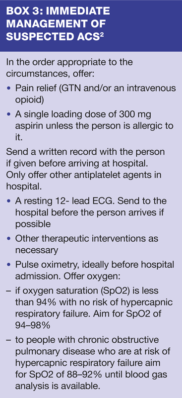
If the patient presents with symptoms suggestive of ACS and they are still experiencing chest pain, immediate urgent care should be instigated and urgent admission arranged.2 (Box 3). The acronym MONA - morphine, oxygen, nitrate (GTN) and aspirin 300mg - is sometime used to describe immediate care, although NICE cautions about the indiscriminate use of oxygen.
Initial assessment
The NICE guideline states that in an acute presentation of chest pain where cardiac causes are suspected the most important initial investigation is an ECG.2 However, studies have shown that a single ECG can provide limited or even misleading information.13 An initial ECG may fail to show any changes, even when the patient is subsequently diagnosed with ACS. For this reason, serial ECG may be more effective as this is more likely to show changes as they develop. These changes might include ST elevation, left bundle branch block or the presence of Q waves2 and may be seen in the ambulance on the way to hospital, in the A&E department or even later.14 It is important to bear in mind that the initial ECG at presentation is not always diagnostic of ACS. The NICE guideline therefore stipulates that if a diagnosis of ACS is suspected, admission should not be delayed pending, or on the basis of an ECG. Other tests, such as troponin T levels,14 can then be carried out in hospital.
FURTHER ASSESSMENT
If ACS is not suspected then other causes for chest pain, which might still include life-threatening conditions, such as pulmonary embolism, should be considered. Once a thorough history has been taken the next step is to carry out further assessment, including examination of the patient and appropriate, objective tests, to help support or refute any tentative diagnosis that has been made.
The history should have given some indication as to the likely cause of the pain, whether the cause is an acute or non-acute problem, and also whether the pain is likely to be cardiac or not. This information will dictate how the rest of the consultation and ongoing care is carried out. Examination may include standard checks such as temperature, pulse rate and rhythm, blood pressure and respiratory rate along with observation of the patient for evidence of obesity, expressions of pain, sweating, nausea and/or vomiting, cough, sputum and shortness of breath.15 Examination skills which may be implemented by a competent and appropriately trained practitioner to identify other causes of chest pain could include respiratory and cardiovascular examination, such as auscultation of the lung and/or heart sounds, assessment of finger clubbing or cyanosis, measurement of the jugular venous pressure (JVP) and abdominal examination for swelling, tenderness or possible organomegaly.
STABLE CHEST PAIN
The NICE guideline on assessing chest pain2 advises that the diagnosis of stable angina can be made on the basis of the clinical assessment, with or without diagnostic testing.
The typical feature of angina is pain that is:
- Constricting and felt in the front of the chest, neck, shoulders, jaw or arms
- Provoked by activity
- Relieved by rest or GTN within about 5 minutes.
People with typical angina will have all of these features; atypical angina presents with two. People with none or only one of these features are said to have non-anginal pain.
The NICE guideline includes a table for assessing the probability that a patient has CHD, based on these features plus the age and sex of the patient along with other factors which increase the risk of pain being due to coronary artery disease, such as diabetes, smoking and hyperlipidaemia. The urgency of further investigations at this stage will depend on the score and on local guidelines. Both the Scottish Intercollegiate Guidelines Network (SIGN)5 and NICE guidelines2 recommend that if a diagnosis of non-ACS CHD is suspected, referral to the Rapid Access Chest Pain (RACP) clinic will help to confirm or refute the diagnosis. This can help minimise unnecessary anxiety for both the patient and their family. The RACP clinic can provide further, specialist input.16
NON-CARDIAC CHEST PAIN
According to NICE,2 features that make a diagnosis of cardiac chest pain less likely, are when the pain is:
- Continuous or very prolonged, and/or
- Unrelated to activity, and/or
- Brought on by breathing in, and/or
- Associated with symptoms such as dizziness, palpitations, tingling or difficulty swallowing.
When patients present with symptoms such as these, NICE recommends that they and their family should be advised that the cause of their pain is not thought to be cardiac and that other potential causes of chest pain are being considered. Common causes of non-cardiac chest pain presenting in primary care include:
- Musculoskeletal pain
- Gastrointestinal disorders
- Anxiety.17
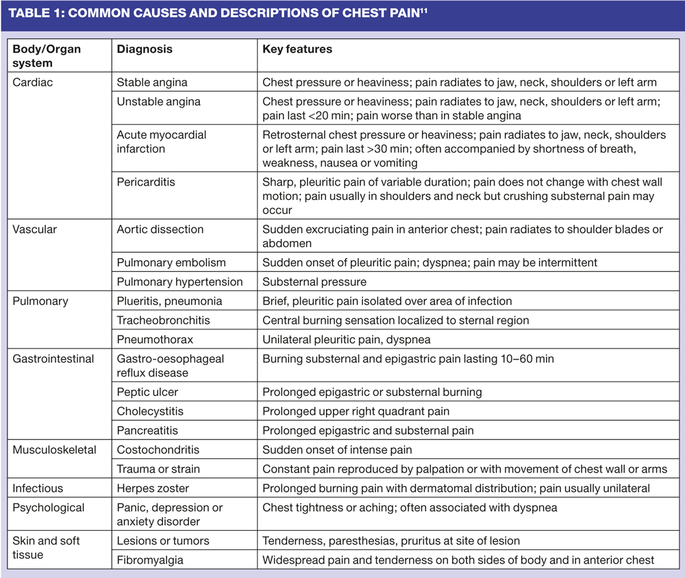
Musculoskeletal pain is often reproduced by pressure; gastrointestinal pain may be linked to food intake. Other possible causes of non-cardiac pain include chest infections, such as pneumonia, which may present with pyrexia, malaise, cough as well as chest or abdominal pain.18 (Table 1)
In non-acute pain, further investigations such as blood tests can be carried out to identify possible causes. These may include:
- Full blood count for anaemia or evidence of infection
- Renal, liver and thyroid function to look for evidence of underlying disease
- Glucose levels which may identify undiagnosed diabetes
- Lipid levels as a risk factor for cardiovascular disease.
Depending on the findings so far, other investigations may be indicated. For example, if the history points towards a possible diagnosis of peptic ulcer, a helicobacter pylori stool or breath test may be requested; if gall stones are suspected an ultrasound scan of the liver and gall bladder may be indicated; chest X-ray or CT may be helpful if lung pathology is suspected. In addition to identifying CHD, an ECG may help in the diagnosis of cardiac arrhythmia or ventricular hypertrophy. There are a multitude of options available, underlining the importance of taking a thorough history to guide appropriate use of further investigative tests and referrals.
CONCLUSION
In summary, chest pain is a common presentation in UK general practices, but most will have a non-cardiac cause. A careful and thorough history will identify people who are acutely unwell and those whose symptoms are more chronic in nature. It is important to be able to quickly identify those patients who have acute chest pain suggestive of ACS so that they can be admitted as an emergency. In those people who have symptoms which are more in line with a diagnosis of stable angina, further calculation of the likelihood of them having CHD can be made using score charts. Diagnostic investigation can then be carried out, if necessary, and the patient can be given appropriate treatment for a diagnosis of CHD. For the majority of people who have non-cardiac chest pain, further assessment and diagnosis will help to confirm the cause of the pain and allow ongoing management based on accurate diagnosis.
REFERENCES
1. O'Toole L. Angina (stable). Clin Evid 2005;13:62-9
2. NICE. Chest pain of recent onset: quick reference guide. NICE 2010. CG95 http://guidance.nice.org.uk/CG95/QuickRefGuide/pdf/English
3. University of Central London Guide to History Taking and Examination http://www.ucl.ac.uk/pcph/undergrad/cbt/year3/history-and-examination
4. British Heart Foundation. Heart disease facts. 2012
http://www.bhf.org.uk/media/news-from-the-bhf/bhf-facts.aspx
5. Scottish Intercollegiate Guidelines Network. Management of stable angina. A national clinical guideline. Edinburgh. SIGN. 2007. Guideline 96
6. Department of Health. Supplementary Prescribing. 2005 http://www.dh.gov.uk/en/Publicationsandstatistics/Publications/PublicationsPolicyAndGuidance/DH_4110032
7. Bickley LS. Guide to Physical Examination and History Taking. Lippincott Williams & Wilkins. 2008
8. Roche B (1998) Assessment by PQRST checklist. 1998 http://www.wemsi.org/pqrst.html
9. Clayton HA. SOCRATES on pain assessment. 2000. Cited in eNotes. 2012 Available from http://www.enotes.com/topic/Socrates_%28pain_assessment%29
10. Rushforth H. Assessment made incredibly easy. Lippincott, Williams & Wilkins. 2009
11. Gibson JL. Evaluating and Treating Chest Pain in the Acute Care Setting US Pharm 2011; 36(5):HS-15-HS-26. Available from http://www.uspharmacist.com/content/d/feature/i/1500/c/28122/
12. Eden BM. Chest pain in women: What's the difference? Nurse Pract 2008;33(2):24-34.
13. Snow V, Barry P, Fihn SD et al. Evaluation of primary care patients with chronic stable angina: guidelines from the American College of Physicians. Ann Intern Med 2004;141:57-64
14. Achar SA, Kundu S and Norcross WA. Diagnosis of Acute Coronary Syndrome Am Fam Physician 2005; 72(01):119-126.
15. Douglas G, Nicol F, Robertson C. Macleod's Clinical Examination. 12th Edition. Churchill Livingstone Elsevier. 2009
16. Keenan J (date not available) Rapid Access Chest Pain Clinic - an update Powerpoint presentation Available from http://www.oxfordlearning.org.uk/oxfordprimarycarelearning/P/Presentations/pdf/R/Rapid%20Access%20Chest%20Pain%20Clinic_%20an%20Update%20%28Jan%20Keenan%29.pdf
17. Cayley WE Jr. Diagnosing the cause of chest pain. Am Fam Physician 2005;72(10):2012-21
18. NHS Choices. Chest Pain. 2011 Available from http://www.nhs.uk/conditions/chest-pain/Pages/Introduction.aspx

Related articles
View all Articles